. 2021 Apr;11(4):200390.
doi: 10.1098/rsob.200390. Epub 2021 Apr 28.

Ubiquitin-proteasome system and the role of its inhibitors in cancer therapy

Affiliations

Ubiquitin-proteasome system and the role of its inhibitors in cancer therapy

Fatemeh Aliabadi et al. Open Biol. 2021 Apr.

Abstract

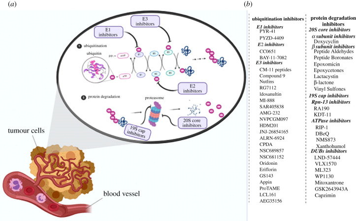
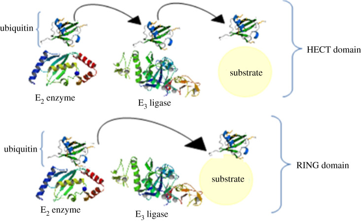
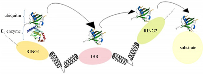
Despite all the other cells that have the potential to prevent cancer development and metastasis through tumour suppressor proteins, cancer cells can upregulate the ubiquitin-proteasome system (UPS) by which they can degrade tumour suppressor proteins and avoid apoptosis. This system plays an extensive role in cell regulation organized in two steps. Each step has an important role in controlling cancer. This demonstrates the importance of understanding UPS inhibitors and improving these inhibitors to foster a new hope in cancer therapy. UPS inhibitors, as less invasive chemotherapy drugs, are increasingly used to alleviate symptoms of various cancers in malignant states. Despite their success in reducing the development of cancer with the lowest side effects, thus far, an appropriate inhibitor that can effectively inactivate this system with the least drug resistance has not yet been fully investigated. A fundamental understanding of the system is necessary to fully elucidate its role in causing/controlling cancer. In this review, we first comprehensively investigate this system, and then each step containing ubiquitination and protein degradation as well as their inhibitors are discussed. Ultimately, its advantages and disadvantages and some perspectives for improving the efficiency of these inhibitors are discussed.

Keywords: cancer; protein degradation inhibitors; targeted therapy; ubiquitination inhibitors; ubiquitin–proteasome system.


Figures

Figure 1.
Figure 1.
The 20S CP structure and its subunits. 20S complexes have four heptameric rings with two separate subunits: α and β subunits. α subunits are located in two ends of the proteasome core. Two inner rings are formed from seven β subunits in which β1, β2 and β3 act as caspase, trypsin and chymotrypsin, respectively.
Figure 2.
Figure 2.
Structure of the 19S regulatory cap. The 19S regulatory cap is divided into two subunits: a base with nine proteins (Rpt1–6, Rpn1, 2, 13) in which six of them are ATPase and directly connected to the α subunit, and a lid with 10 proteins (Rpn3, 5–12, 15).
Figure 3.
Figure 3.
Ubiquitination process. First, E1 hydrolyses ATP and then adenylates one ubiquitin molecule. Later, this ubiquitin is translocated to the cysteine active site of E1. After that, this adenylated ubiquitin is translocated to the cysteine of E2. Finally, E3 recognizes the targeted protein and catalyses the translocation of the ubiquitin from E2 to the protein.
Figure 4.
Figure 4.
(a) Ubiquitin-proteasome system inhibitors. (b) List of the inhibitors of the ubiquitination process as well as protein degradation.
Figure 5.
Figure 5.
Two main classes of E3 ligases. HECT ligases have a C-terminal domain that accepts a ubiquitin molecule from the E2 enzyme by the formation of a thioester bond before the ubiquitin translocation to the substrate, and RING ligases, which have a zinc finger letting E2 directly transfer the ubiquitin to the substrate.
Figure 6.
Figure 6.
RING1-IBR (in-between-RING)-RING2 ubiquitin ligase. RBR (RING-between-RING) has two domains having elements of both RING and HECT ligases; one RING domain binds to the E2 while another RING domain accepts the ubiquitin molecule before its translocation to the substrate.
Figure 7.
Figure 7.
Comparison between the inhibition activity of Bortezomib and shPSMA1 on chymotrypsin proteasome. PSMA1 knockdown showed more inhibition compared to Bortezomib. CTL, chymotrypsin-like; Veh, vehicle. Adapted from [184]. Copyright © 2013 Public Library of Science.
Figure 8.
Figure 8.
The process of protein proteolysis by chymotrypsin. (a) The substrate becomes acylated to form an acyl-enzyme intermediate. The hydrogen of the aspartate binds to the N-δ hydrogen of histidine. (b) The sidelong chain of the serine acts as a nucleophile and binds to the carbonyl carbon of the substrate's main chain. (c) Ionization of carbonyl oxygen becomes stabilized by the formation of two sidelong hydrogen bonds to the N-hydrogens of the main chain. These reactions result in a tetrahedral combination and cause the peptide bond to be broken down. (d) Formation of an acyl-enzyme intermediate bonded to serine and causes the new N-terminal of the protein to be broken down and separated. Moreover, in the second step of the reaction, one water molecule becomes activated by histidine and acts as a nucleophile. The oxygen of the water attacks the carbonyl carbon of the acyl group of serine, which results in the formation of a secondary tetrahedral combination, regeneration of the OH group of serine and the release of a proton as well as a protein with a newly formed C-terminal domain.
Figure 9.
Figure 9.
The suggested mechanism for the reaction of epoxomicin (EPX) with proteasomes. Possible mechanisms for this reaction are via five steps. (i) Proton transfer process, activating Thr1-Oγ directly via Thr1-Nz to form a zwitterionic intermediate. (ii) Nucleophilic attack on the carbonyl carbon of EPX by the negatively charged Thr1-Oγ atom. (iii) Proton transfer from Thr1-Nz to the carbonyl oxygen of EPX. (iv) Thr1-Nz attacks the carbon of the epoxide group of EPX, along with the epoxide ring-opening (SN2 nucleophilic substitution) so that a zwitterionic morpholino ring is formed between residue Thr1 and EPX. (v) The product of the morpholino ring is generated by another proton transfer. Adapted from [196]. Copyright © 2012 American Chemical Society.

References

    1. Varshavsky A. 2006. The early history of the ubiquitin field. Protein Sci. 15, 647-654. (10.1110/ps.052012306) - DOI - PMC - PubMed
    1. Hershko A, Ciechanover A, Heller H, Haas AL, Rose IA. 1980. Proposed role of ATP in protein breakdown: conjugation of protein with multiple chains of the polypeptide of ATP-dependent proteolysis. Proc. Natl Acad. Sci. USA 77, 1783-1786. (10.1073/pnas.77.4.1783) - DOI - PMC - PubMed
    1. Hershko A, Heller H, Elias S, Ciechanover A. 1983. Components of ubiquitin–protein ligase system. Resolution, affinity purification, and role in protein breakdown. J. Biol. Chem. 258, 8206-8214. (10.1016/S0021-9258(20)82050-X) - DOI - PubMed
    1. Hershko A, Ciechanover A, Varshvsky A. 2000. The ubiquitin system. Nat. Med. 10, 1073-1081. (10.1038/80384) - DOI - PubMed
    1. Giles RE, Blanc H, Cann HM, Wallace DC. 1980. Maternal inheritance of human mitochondrial DNA. Proc. Natl Acad. Sci. USA 77, 6715-6719. (10.1073/pnas.77.11.6715) - DOI - PMC - PubMed

MeSH terms

LinkOut - more resources