Review
. 2021 Apr 27;21(1):239.
doi: 10.1186/s12935-021-01946-4.

Small molecule inhibitors against PD-1/PD-L1 immune checkpoints and current methodologies for their development: a review

Affiliations
Review

Small molecule inhibitors against PD-1/PD-L1 immune checkpoints and current methodologies for their development: a review

Chang Liu et al. Cancer Cell Int. .

Abstract

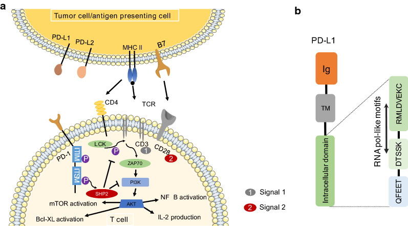
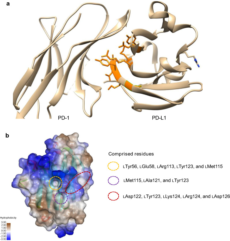
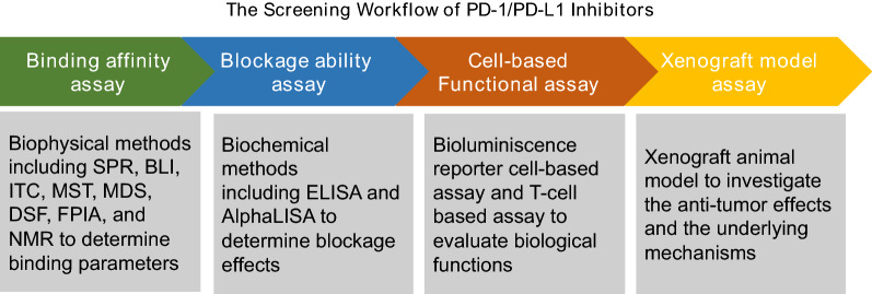
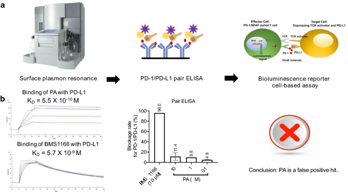
Programmed death-1/programmed death ligand-1 (PD-1/PD-L1) based immunotherapy is a revolutionary cancer therapy with great clinical success. The majority of clinically used PD-1/PD-L1 inhibitors are monoclonal antibodies but their applications are limited due to their poor oral bioavailability and immune-related adverse effects (irAEs). In contrast, several small molecule inhibitors against PD-1/PD-L1 immune checkpoints show promising blockage effects on PD-1/PD-L1 interactions without irAEs. However, proper analytical methods and bioassays are required to effectively screen small molecule derived PD-1/PD-L1 inhibitors. Herein, we summarize the biophysical and biochemical assays currently employed for the measurements of binding capacities, molecular interactions, and blocking effects of small molecule inhibitors on PD-1/PD-L1. In addition, the discovery of natural products based PD-1/PD-L1 antagonists utilizing these screening assays are reviewed. Potential pitfalls for obtaining false leading compounds as PD-1/PD-L1 inhibitors by using certain binding bioassays are also discussed in this review.

Keywords: Cancer; Immunotherapy; Natural products; PD-1/PD-L1; Small molecules.


Conflict of interest statement

The authors declare no competing financial interests.

Figures

Fig. 1
Fig. 1
The signaling pathway of PD-1/PD-L1. a PD-L1 consists of an extracellular domain, a transmembrane domain, and an intracytoplasmic region but lacks intracellular signaling. The intracytoplasmic region consists of three conserved sequences including RMLDVEKC, DTSSK, and QFEET motifs. The part of the RMLDVEKC motif and the entire DTSSK motif that have been identified by MotifFinder are RNA pol-like motifs. b Antigens are presented by APCs as antigenic peptides, which are recognized by the T-cell receptor (TCR; Signal 1). The second signal (Signal 2) is delivered when B7 (CD80 and CD86) on the APCs engage CD28 on the T cells
Fig. 2
Fig. 2
The structure of PD-L1 (4ZQK) and three main hot spots between PD-1 and PD-L1. a The structures of PD-L1 and PD-1. Amino acid residues in the main hot spots are labeled as orange color. b Three main hot spots are exhibited. The first hotspot includes lTyr56, lGlu58, lArg113, lMet115, and lTyr123. The second hotspot includes lMet115, lAla121, and lTyr123. The third hotspot is an extended groove formed by the main chain and the side chains spanning residues lAsp122 to lArg125, and is flanked by the side chain of lAsp26
Fig. 3
Fig. 3
The screening workflow of PD-1/PD-L1 inhibitors. The identification of PD-1/PD-L1 inhibitors is required by using a series of assays including binding affinity assay, blockage ability assay, cell-based functional assay and xenograft model assay
Fig. 4
Fig. 4
Chemical structures of natural products based PD-1/PD-L1 inhibitors including cyclo(-Leu-DTrp-Pro-Thr-Asp-Leu-DPhe-Lys(Dde)-Val-Arg-), rifabutin, kaempferol, kaempferol-7-O-rhamnoside, eriodictyol, fisetin, glyasperin C, cosmosiin, ellagic acid, and caffeoylquinic acids
Fig. 5
Fig. 5
A workflow was established for screening PD-1/PD-L1 inhibitors. a SPR technology was performed to evaluate binding affinities followed by PD-1/PD-L1 pair ELISA assay. Once the inhibitors exert blockage effects on PD-1/PD-L1 interactions, bioluminescence reporter cell-based assay will be applied for determining their biological functions. b The binding and inhibitory effects of Punicalagin (PA) and BMS1166 against human PD-L1 protein assessed using SPR and PD-1/PD-L1 pair ELISA assays, respectively. PA or BMS1166 was allowed to flow over Fc-PD-L1 captured on a flow cell as well as on a reference cell of Series S Sensor Chip

References

    1. Fernandes M, Brabek J. Cancer, checkpoint inhibitors, and confusion. Lancet Oncol. 2017;18(11):e632. doi: 10.1016/S1470-2045(17)30810-0. - DOI - PubMed
    1. Dunn GP, Bruce AT, Ikeda H, Old LJ, Schreiber RD. Cancer immunoediting: from immunosurveillance to tumor escape. Nat Immunol. 2002;3(11):991–998. doi: 10.1038/ni1102-991. - DOI - PubMed
    1. Sathyanarayanan V, Neelapu SS. Cancer immunotherapy: strategies for personalization and combinatorial approaches. Mol Oncol. 2015;9(10):2043–2053. doi: 10.1016/j.molonc.2015.10.009. - DOI - PMC - PubMed
    1. Durant JR. Immunotherapy of cancer: the end of the beginning? N Engl J Med. 1987;316(15):939–941. doi: 10.1056/NEJM198704093161509. - DOI - PubMed
    1. Farkona S, Diamandis EP, Blasutig IM. Cancer immunotherapy: the beginning of the end of cancer? BMC Med. 2016;14:73. doi: 10.1186/s12916-016-0623-5. - DOI - PMC - PubMed