

COVID-19 triggering mucormycosis in a susceptible patient: a new phenomenon in the developing world?
- PMID: 33906877
- PMCID: PMC8088249
- DOI: 10.1136/bcr-2021-241663
COVID-19 triggering mucormycosis in a susceptible patient: a new phenomenon in the developing world?
Abstract
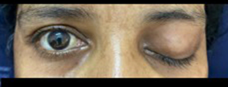
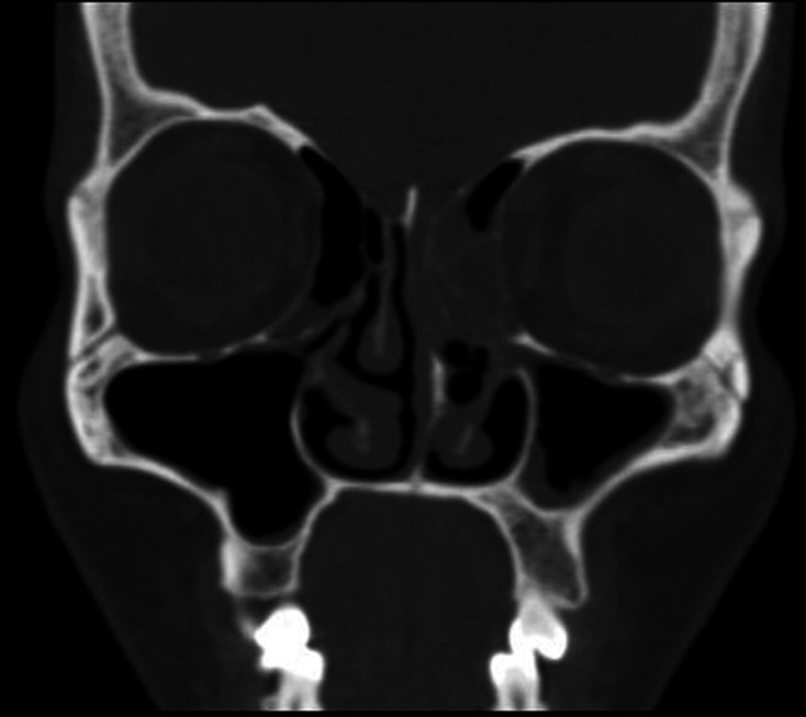
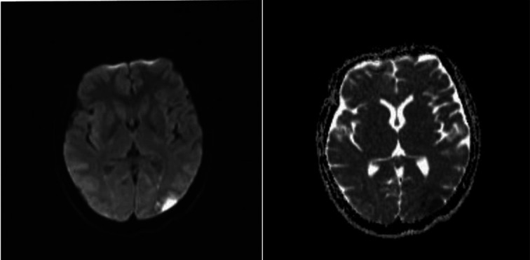
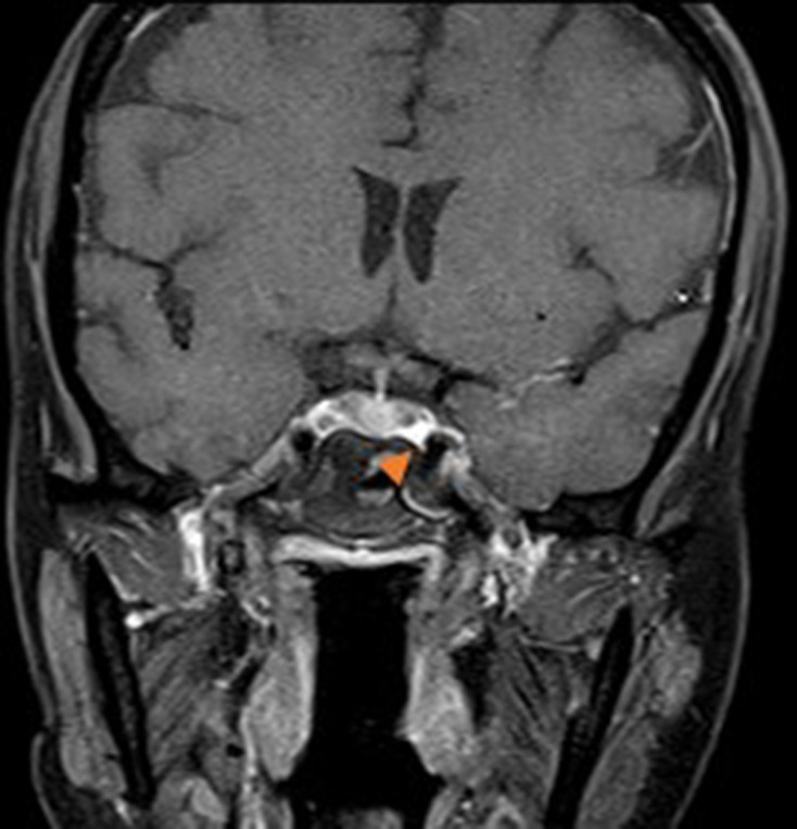
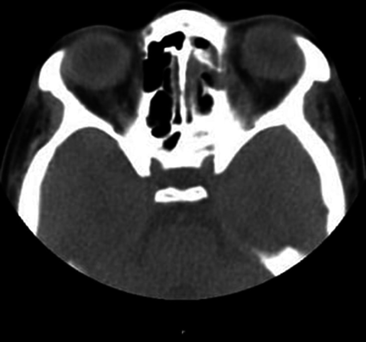
A middle-aged woman with diabetes presented with left-sided facial pain, complete ptosis and fever of short duration. On presentation, she had hyperglycaemia without ketosis. There was total ophthalmoplegia of the left eye with a visual acuity of 6/36. She incidentally tested positive for COVID-19. CT paranasal sinus and MRI brain revealed left-sided pansinusitis with acute infarct in the left parieto-occipital region without angioinvasion. An emergency functional endoscopic sinus procedure was done, which confirmed mucormycosis on histopathological examination. After 1 week of conventional amphotericin B and antibiotics, repeat CT brain showed improvement in mucosal thickening and sinusitis. This case is a rare presentation of mucormycosis associated with rapid progression to orbital apex syndrome with brain infarction in a patient with non-ketotic diabetes and COVID-19. Early diagnosis and treatment are essential to prevent further end-organ damage. It is also interesting that there was no angioinvasion and transient periarterial inflammation was attributed to brain infarction.
Keywords: COVID-19; diabetes; stroke; tropical medicine (infectious disease).
© BMJ Publishing Group Limited 2021. No commercial re-use. See rights and permissions. Published by BMJ.
Conflict of interest statement
Competing interests: None declared.
Figures
Comment in
-
Mucormycosis Epidemic and Stroke in India During the COVID-19 Pandemic.Stroke. 2021 Oct;52(10):e622-e623. doi: 10.1161/STROKEAHA.121.036626. Epub 2021 Sep 16. Stroke. 2021. PMID: 34525840 No abstract available.
References
Publication types
MeSH terms
Substances
LinkOut - more resources
Full Text Sources
Medical
Miscellaneous