. 2021 Apr 27;12(2):e00085-21.
doi: 10.1128/mBio.00085-21.

Analysis of the Long-Term Impact on Cellular Immunity in COVID-19-Recovered Individuals Reveals a Profound NKT Cell Impairment

Affiliations

Analysis of the Long-Term Impact on Cellular Immunity in COVID-19-Recovered Individuals Reveals a Profound NKT Cell Impairment

Jia Liu et al. mBio. .

Abstract

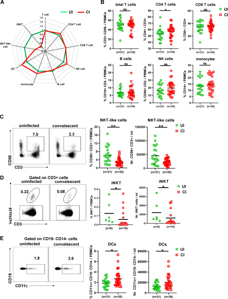
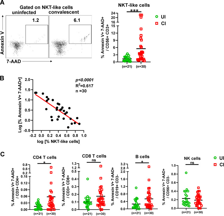
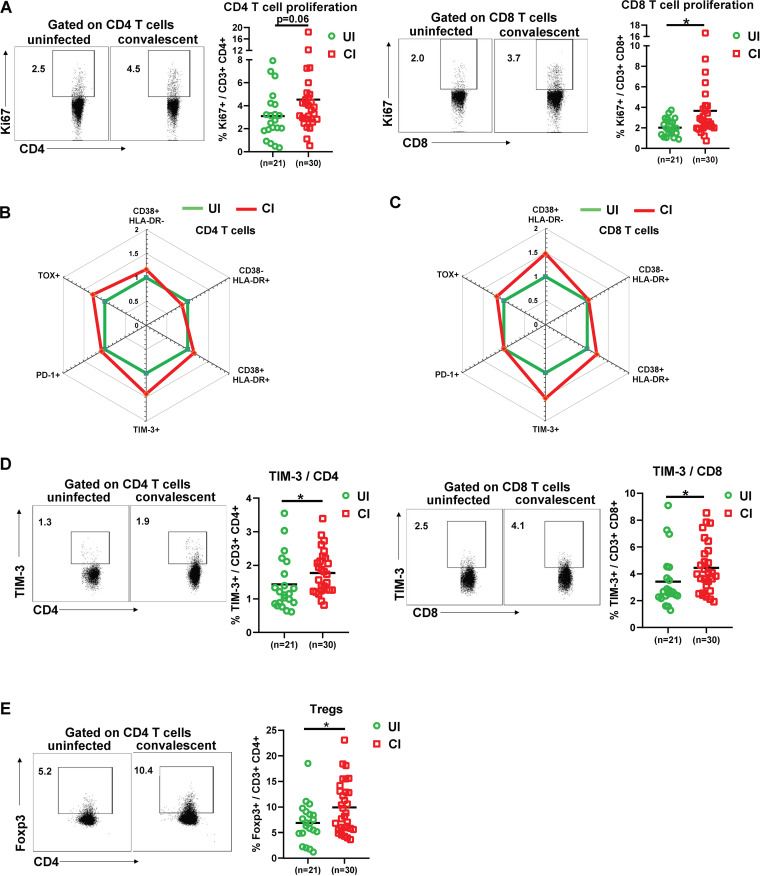
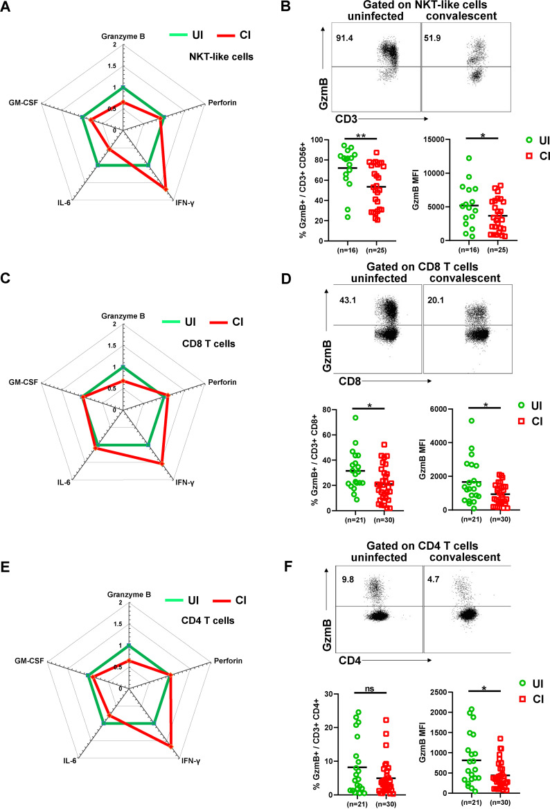
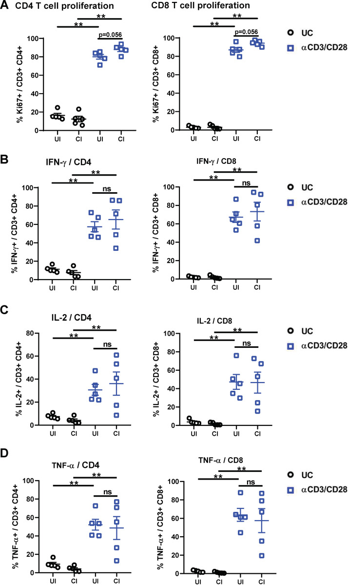
The coronavirus disease 2019 (COVID-19) pandemic caused by the severe acute respiratory syndrome coronavirus 2 (SARS-CoV-2) affected over 120 million people and killed over 2.7 million individuals by March 2021. While acute and intermediate interactions between SARS-CoV-2 and the immune system have been studied extensively, long-term impacts on the cellular immune system remain to be analyzed. Here, we comprehensively characterized immunological changes in peripheral blood mononuclear cells in 49 COVID-19-convalescent individuals (CI) in comparison to 27 matched SARS-CoV-2-unexposed individuals (UI). Despite recovery from the disease for more than 2 months, CI showed significant decreases in frequencies of invariant NKT and NKT-like cells compared to UI. Concomitant with the decrease in NKT-like cells, an increase in the percentage of annexin V and 7-aminoactinomycin D (7-AAD) double-positive NKT-like cells was detected, suggesting that the reduction in NKT-like cells results from cell death months after recovery. Significant increases in regulatory T cell frequencies and TIM-3 expression on CD4 and CD8 T cells were also observed in CI, while the cytotoxic potential of T cells and NKT-like cells, defined by granzyme B (GzmB) expression, was significantly diminished. However, both CD4 and CD8 T cells of CI showed increased Ki67 expression and were fully able to proliferate and produce effector cytokines upon T cell receptor (TCR) stimulation. Collectively, we provide a comprehensive characterization of immune signatures in patients recovering from SARS-CoV-2 infection, suggesting that the cellular immune system of COVID-19 patients is still under a sustained influence even months after the recovery from disease.IMPORTANCE Wuhan was the very first city hit by SARS-CoV-2. Accordingly, the patients who experienced the longest phase of convalescence following COVID-19 reside here. This enabled us to investigate the "immunological scar" left by SARS-CoV-2 on cellular immunity after recovery from the disease. In this study, we characterized the long-term impact of SARS-CoV-2 infection on the immune system and provide a comprehensive picture of cellular immunity of a convalescent COVID-19 patient cohort with the longest recovery time. We revealed that the cellular immune system of COVID-19 patients is still under a sustained influence even months after the recovery from disease; in particular, a profound NKT cell impairment was found in the convalescent phase of COVID-19.

Keywords: COVID-19; NKT cell; SARS-CoV-2; cellular immunity.


Figures

FIG 1
FIG 1
Characterization of immune cell subsets in individuals recovering from COVID-19. (A) The fold changes of the percentages (median) of total T cells, CD4 and CD8 T cells, B cells, NK cells, NKT-like cells, dendritic cells, and monocytes in the blood of CI (n = 30) compared to those of UI (n = 21) are depicted by radar plots. (B) The percentages of total T cells, CD4 and CD8 T cells, B cells, NK cells, and monocytes in the blood of UI (n = 21) and CI (n = 30) were analyzed by flow cytometry. (C) The absolute numbers and percentages of NKT-like cells in the blood of UI (n = 21) and CI (n = 30) were analyzed by flow cytometry. (D) The absolute numbers and percentages of invariant NKT (iNKT) cells in the blood of UI (n = 6) and CI (n = 19) were analyzed by flow cytometry. (E) The absolute numbers and percentages of dendritic cells in the blood of UI (n = 21) and CI (n = 30) were analyzed by flow cytometry. CI, COVID-19-convalescent individuals; UI, SARS-CoV-2-unexposed individuals. Statistically significant differences are indicated by asterisks (*, <0.05; **, <0.01; nonparametric Mann-Whitney test).
FIG 2
FIG 2
Characterization of immune cell death in individuals recovering from COVID-19. (A) The percentages of early apoptosis (annexin V+ 7-AAD) and late apoptosis/necroptosis (annexin V+ 7-AAD+) of NKT-like cells in the blood of CI (n = 30) compared to those of UI (n = 21) were analyzed by flow cytometry. (B) Correlation analysis between the frequencies and the late apoptosis/necroptosis of NKT-like cells was performed in CI. (C) The percentages of early apoptosis (annexin V+ 7-AAD) and late apoptosis/necroptosis (annexin V+ 7-AAD+) of CD4 T, CD8 T, B, and NK cells in the blood of CI (n = 30) compared to those of UI (n = 21) were analyzed by flow cytometry. CI, COVID-19-convalescent individuals; UI, SARS-CoV-2-unexposed individuals. Statistically significant differences are indicated by asterisks (*, <0.05; **, <0.01; ns, not significant; nonparametric Mann-Whitney test).
FIG 3
FIG 3
Characterization of T cell phenotypes in the PBMCs of individuals recovering from COVID-19. (A) The percentages of Ki67+ CD4 and CD8 T cells in the blood of UI (n = 21) and CI (n = 30) were analyzed by flow cytometry. (B and C) The fold changes of the percentages (median) of CD38+ HLA-DR, CD38 HLA-DR+, CD38+ HLA-DR+, PD-1+ TOX, PD-1 TOX+, PD-1+ TOX+, and TIM-3+ CD4 and CD8 T cells in the blood of CI (n = 30) compared to those of UI (n = 21) are depicted by radar plots. (D) The percentages of TIM-3+ CD4 and CD8 T cells in the blood of UI (n = 21) and CI (n = 30) were analyzed by flow cytometry. (E) The percentages of Foxp3+ CD4 T cells in the blood of UI (n = 21) and CI (n = 30) were analyzed by flow cytometry. CI, COVID-19-convalescent individuals; UI, SARS-CoV-2-unexposed individuals. Statistically significant differences are indicated by asterisks (*, <0.05; **, <0.01; nonparametric Mann-Whitney test).
FIG 4
FIG 4
Characterization of cytotoxic and cytokine profiles of T, NK, and NKT-like cells in the PBMCs of individuals recovering from COVID-19. (A) The fold changes of the percentages (median) of granzyme B-, perforin-, IFN-γ-, IL-6-, and GM-CSF-producing NKT-like cells in the blood of CI compared to those of UI are depicted by radar plots. (B) The percentages and MFI (geometric mean) of granzyme B expression of NKT-like cells in the blood of UI (n = 16) and CI (n = 25) were analyzed by flow cytometry. (C) The fold changes of the percentages (median) of granzyme B-, perforin-, IFN-γ-, IL-6-, and GM-CSF-producing CD8 T cells in the blood of CI compared to those of UI are depicted by radar plots. (D) The percentages and MFI (geometric mean) of granzyme B expression of CD8 T cells in the blood of UI (n = 21) and CI (n = 30) were analyzed by flow cytometry. (E) The fold changes of the percentages (median) of granzyme B-, perforin-, IFN-γ-, IL-6-, and GM-CSF-producing CD4 T cells in the blood of CI compared to those of UI are depicted by radar plots. (F) The percentages and MFI (geometric mean) of granzyme B expression of CD4 T cells in the blood of UI (n = 21) and CI (n = 30) were analyzed by flow cytometry. CI, COVID-19-convalescent individuals; UI, SARS-CoV-2-unexposed individuals. Statistically significant differences are indicated by asterisks (*, <0.05; **, <0.01; nonparametric Mann-Whitney test).
FIG 5
FIG 5
Characterization of the effector function of CD4 and CD8 T cells in the PBMCs of individuals recovering from COVID-19. PBMCs of UI (n = 5) and CI (n = 5) were either stimulated with anti-CD3 and anti-CD28 antibodies (αCD3/CD28) or left unstimulated (UC) and cultured for 5 days. The percentages of Ki67 (A)-, IFN-γ (B)-, IL-2 (C)-, and TNF-α (D)-positive CD4 (left) and CD8 (right) T cells were analyzed by flow cytometry. CI, COVID-19-convalescent individuals; UI, SARS-CoV-2-unexposed individuals. Statistically significant differences are indicated by asterisks (*, <0.05; **, <0.01; nonparametric Mann-Whitney test).

Similar articles

Cited by

References

    1. Long QX, Tang XJ, Shi QL, Li Q, Deng HJ, Yuan J, Hu JL, Xu W, Zhang Y, Lv FJ, Su K, Zhang F, Gong J, Wu B, Liu XM, Li JJ, Qiu JF, Chen J, Huang AL. 2020. Clinical and immunological assessment of asymptomatic SARS-CoV-2 infections. Nat Med 26:1200–1204. 10.1038/s41591-020-0965-6. - DOI - PubMed
    1. Guan WJ, Ni ZY, Hu Y, Liang WH, Ou CQ, He JX, Liu L, Shan H, Lei CL, Hui DSC, Du B, Li LJ, Zeng G, Yuen KY, Chen RC, Tang CL, Wang T, Chen PY, Xiang J, Li SY, Wang JL, Liang ZJ, Peng YX, Wei L, Liu Y, Hu YH, Peng P, Wang JM, Liu JY, Chen Z, Li G, Zheng ZJ, Qiu SQ, Luo J, Ye CJ, Zhu SY, Zhong NS, China Medical Treatment Expert Group for Covid-19. 2020. Clinical characteristics of coronavirus disease 2019 in China. N Engl J Med 382:1708–1720. 10.1056/NEJMoa2002032. - DOI - PMC - PubMed
    1. Puelles VG, Lutgehetmann M, Lindenmeyer MT, Sperhake JP, Wong MN, Allweiss L, Chilla S, Heinemann A, Wanner N, Liu S, Braun F, Lu S, Pfefferle S, Schroder AS, Edler C, Gross O, Glatzel M, Wichmann D, Wiech T, Kluge S, Pueschel K, Aepfelbacher M, Huber TB. 2020. Multiorgan and renal tropism of SARS-CoV-2. N Engl J Med 383:590–592. 10.1056/NEJMc2011400. - DOI - PMC - PubMed
    1. Asadi-Pooya AA, Simani L. 2020. Central nervous system manifestations of COVID-19: a systematic review. J Neurol Sci 413:116832. 10.1016/j.jns.2020.116832. - DOI - PMC - PubMed
    1. Huang C, Wang Y, Li X, Ren L, Zhao J, Hu Y, Zhang L, Fan G, Xu J, Gu X, Cheng Z, Yu T, Xia J, Wei Y, Wu W, Xie X, Yin W, Li H, Liu M, Xiao Y, Gao H, Guo L, Xie J, Wang G, Jiang R, Gao Z, Jin Q, Wang J, Cao B. 2020. Clinical features of patients infected with 2019 novel coronavirus in Wuhan, China. Lancet 395:497–506. 10.1016/S0140-6736(20)30183-5. - DOI - PMC - PubMed

Publication types