. 2021 May 4;118(18):e2001681118.
doi: 10.1073/pnas.2001681118.

Genetic removal of p70 S6K1 corrects coding sequence length-dependent alterations in mRNA translation in fragile X syndrome mice

Affiliations

Genetic removal of p70 S6K1 corrects coding sequence length-dependent alterations in mRNA translation in fragile X syndrome mice

Sameer Aryal et al. Proc Natl Acad Sci U S A. .

Abstract

Loss of the fragile X mental retardation protein (FMRP) causes fragile X syndrome (FXS). FMRP is widely thought to repress protein synthesis, but its translational targets and modes of control remain in dispute. We previously showed that genetic removal of p70 S6 kinase 1 (S6K1) corrects altered protein synthesis as well as synaptic and behavioral phenotypes in FXS mice. In this study, we examined the gene specificity of altered messenger RNA (mRNA) translation in FXS and the mechanism of rescue with genetic reduction of S6K1 by carrying out ribosome profiling and RNA sequencing on cortical lysates from wild-type, FXS, S6K1 knockout, and double knockout mice. We observed reduced ribosome footprint (RF) abundance in the majority of differentially translated genes in the cortices of FXS mice. We used molecular assays to discover evidence that the reduction in RF abundance reflects an increased rate of ribosome translocation, which is captured as a decrease in the number of translating ribosomes at steady state and is normalized by inhibition of S6K1. We also found that genetic removal of S6K1 prevented a positive-to-negative gradation of alterations in translation efficiencies (RF/mRNA) with coding sequence length across mRNAs in FXS mouse cortices. Our findings reveal the identities of dysregulated mRNAs and a molecular mechanism by which reduction of S6K1 prevents altered translation in FXS.

Keywords: autism; fragile X syndrome; mRNA translation; protein synthesis; translation elongation.


Conflict of interest statement

The authors declare no competing interest.

Figures
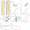
Fig. 1.
Fig. 1.
Increased CDS length is associated with decreased RF abundance and increased mRNA expression in FXS mouse cortices. (A) Heat maps depicting RF abundance (n = 204) and mRNA expression (n = 351) of significantly different genes (FDR-adjusted P value< 0.1) between WT and FXS mouse cortices in ribosome profiling (Left) and RNA-Seq analyses (Right). Each row displays centered and scaled (“row-normalized”) transcripts per million values for significantly different genes. (B) Significance (FDR-adjusted P value) versus LFC in RF abundance between FXS and WT mice. The 20 most significant genes are labeled. (C) Volcano plot of mRNA expression changes in FXS and WT mice. The top 10 significantly different genes and several genes previously shown to be implicated in FXS are labeled. (D) Cumulative distribution of LFCs in RF abundance (FXS/WT) as a function of CDS length (n = 415, 764, 3,494, 5,866, 1,451, and 93 mRNAs in bins 1 through 6). (E) Cumulative distribution of LFCs in mRNA expression (FXS/WT) as a function of CDS length (n = 434, 816, 3,713, 6,252, 1,556, and 105 mRNAs in bins 1 through 6). (F) Top biological processes enriched in FXS and WT mouse cortical tissue in ribosome profiling (upper green-dashed box) or RNA-Seq assays. (G) The translation profile of the Zmiz1 gene. The RF abundance LFC (FXS/WT) and the negative of the mRNA expression LFC (FXS/WT) are added to calculate the LFC in TE. (H) Cumulative distribution of LFCs in translation efficiencies (FXS/WT) as a function of CDS length. Comparison of LFCs in RF abundance and mRNA expression (I) and TE (J) by CDS length in FXS. mRNAs are divided into 50 bins by their CDS lengths. Each bin contains ∼250 mRNAs.
Fig. 2.
Fig. 2.
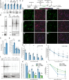
The rate of translation elongation is increased in FXS primary neurons and is sensitive to the inhibition of S6K1. (A) Representative immunoblots and their quantifications examining the protein levels of mRNAs with particularly long (Dst, Herc1, Adcy1, Plec, and Zmiz1) or short (Eif1) CDSs in cortical lysates from WT and FXS mice. (B) Representative Western blot of WT and FXS cortical neurons incubated with puromycin. (C) Quantification of A (t test, P = 0.05). (D) Representative Western blot of the ES assay in WT and FXS neurons. (E) Quantification of C (pairwise t test, holm-adjusted P = 0.007 at baseline). (F) Protocol for ES and runoff-RPM assays. (G) Representative immunofluorescence images of runoff-RPM in WT and FXS mouse cortical cultures. (H) Quantification of G across two experiments. (I) Results from H, expressed as a percentage of its own baseline. (J) Representative Western blot of runoff-RPM assay in WT and FXS mouse neurons treated with the S6K1 inhibitor PF-4708671. (K) Quantification of G. Values are expressed as a percentage of WT + vehicle baseline. (L) The inverse of the rate of runoff elongation. Elongation rate is increased in FXS (t8, WT + vehicle versus FXS + vehicle, holm-adjusted P = 0.005) and is rescued by pharmacological inhibition of S6K1 (pairwise t test, t16 WT + vehicle versus t16 FXS + PF470861 holm-adjusted P = 1). #P < 0.001, **P < 0.01, *P < 0.1, ns: P > 0.1. Pairwise t test used for post hoc comparisons with holm adjustment.
Fig. 3.
Fig. 3.
CDS length dependency of TE and mRNA expression in FXS mouse cortices is corrected by genetic reduction of S6K1. (A) RF profile of Rps6kb1 mRNA. Profile accurately captures the stop codon introduced to make the S6K1 deletion. Dotted lines indicate UTR boundaries. Volcano plot of RF abundance (B) (n = 3) and mRNA expression (C) (n = 4) (DKO/WT). The top 20 genes are labeled. Cumulative distribution of LFCs in RF abundance (D) and mRNA expression (E) in DKO brains, as a function of CDS length. Bin sizes are the same as those used in Fig. 1 D and E, respectively. Comparison of LFCs by CDS length in FXS and DKO brains in mRNA expression (F) and TE (G) datasets. Comparison of RF abundance (H) and mRNA expression (I) in FXS and DKO brains of genes in the longest four bins, compared to the WT. The majority of points are above the x = y line in RF abundance and below the line in RNA expression, suggesting a generalized reduction in RF abundance and mRNA expression toward WT levels. #P < 0.001, **P < 0.01, *P < 0.1. Kruskal–Wallis test used for comparison of LFCs within each bin; P values are holm-adjusted.

References

    1. Qin M., Kang J., Burlin T. V., Jiang C., Smith C. B., Postadolescent changes in regional cerebral protein synthesis: An in vivo study in the FMR1 null mouse. J. Neurosci. 25, 5087–5095 (2005). - PMC - PubMed
    1. Sidorov M. S., Auerbach B. D., Bear M. F., Fragile X mental retardation protein and synaptic plasticity. Mol. Brain 6, 15 (2013). - PMC - PubMed
    1. Darnell J. C., Klann E., The translation of translational control by FMRP: Therapeutic targets for FXS. Nat. Neurosci. 16, 1530–1536 (2013). - PMC - PubMed
    1. Bhattacharya A., et al. ., Genetic removal of p70 S6 kinase 1 corrects molecular, synaptic, and behavioral phenotypes in fragile X syndrome mice. Neuron 76, 325–337 (2012). - PMC - PubMed
    1. Thomson S. R., et al. ., Cell-type-specific translation profiling reveals a novel strategy for treating fragile X syndrome. Neuron 95, 550–563.e5 (2017). - PMC - PubMed

Publication types

Substances

LinkOut - more resources