. 2021 Apr 27;5(4):193-209.
doi: 10.4049/immunohorizons.2100018.

Indole-3-Carbinol-Dependent Aryl Hydrocarbon Receptor Signaling Attenuates the Inflammatory Response in Experimental Necrotizing Enterocolitis

Affiliations

Indole-3-Carbinol-Dependent Aryl Hydrocarbon Receptor Signaling Attenuates the Inflammatory Response in Experimental Necrotizing Enterocolitis

Lila S Nolan et al. Immunohorizons. .

Abstract

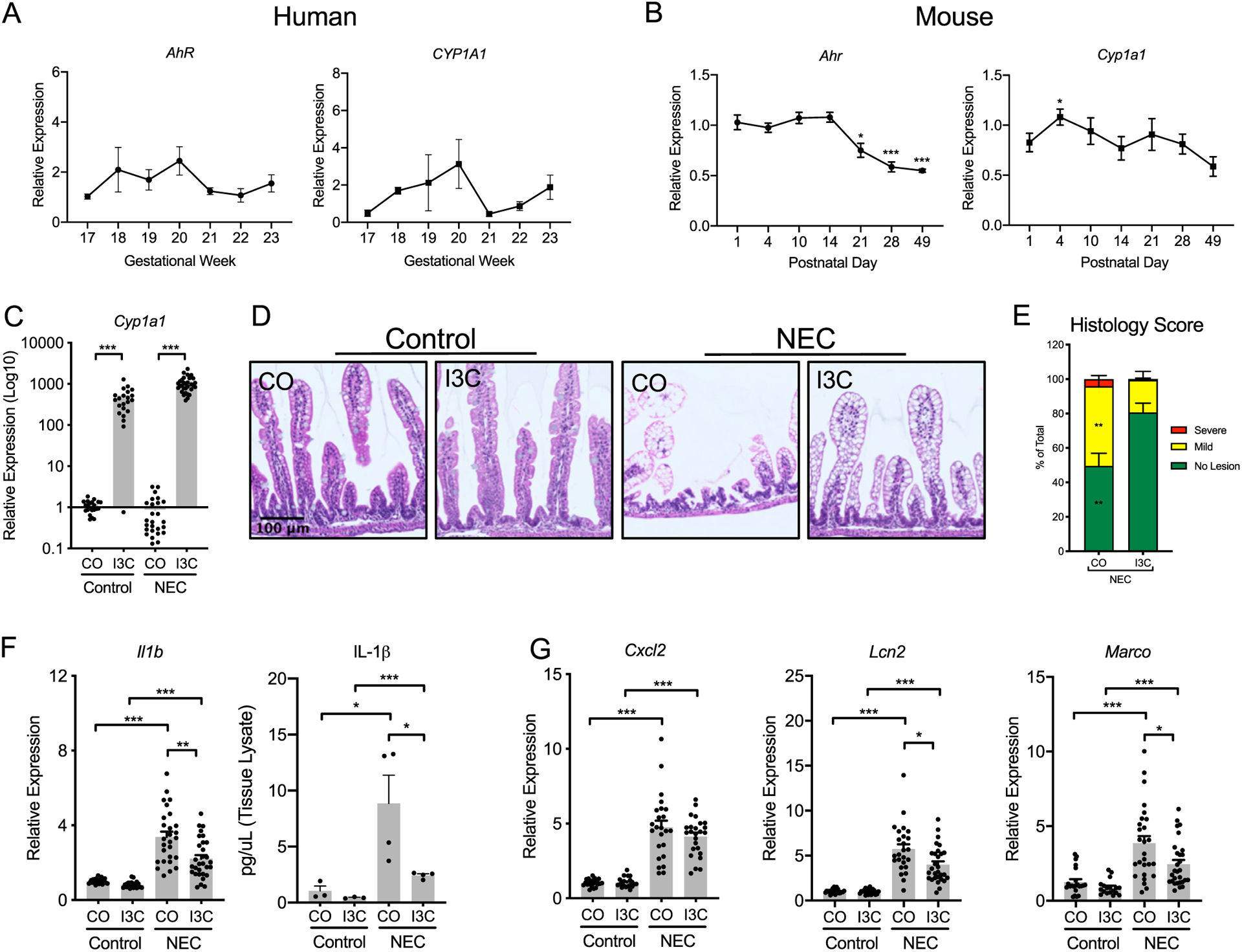
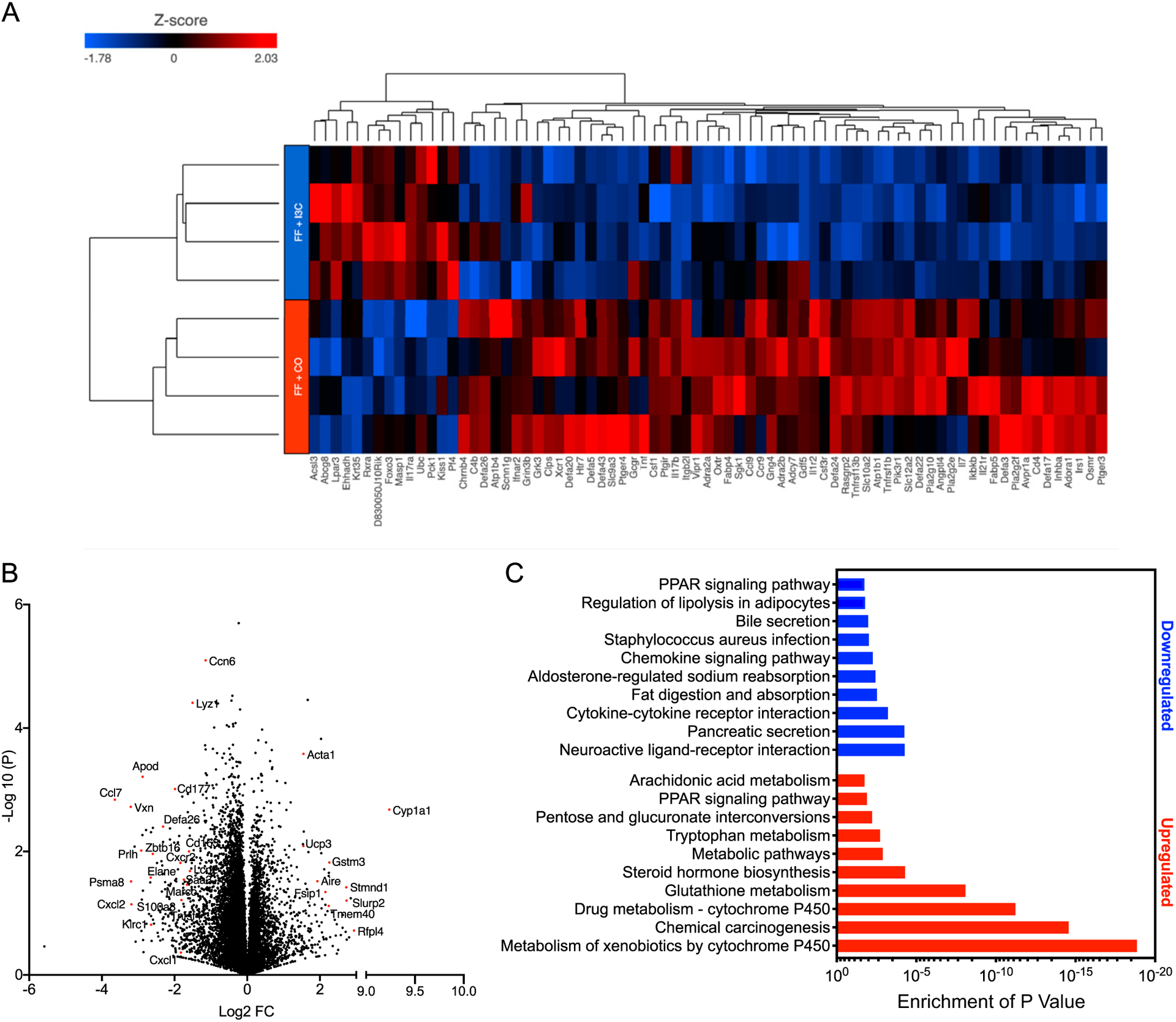
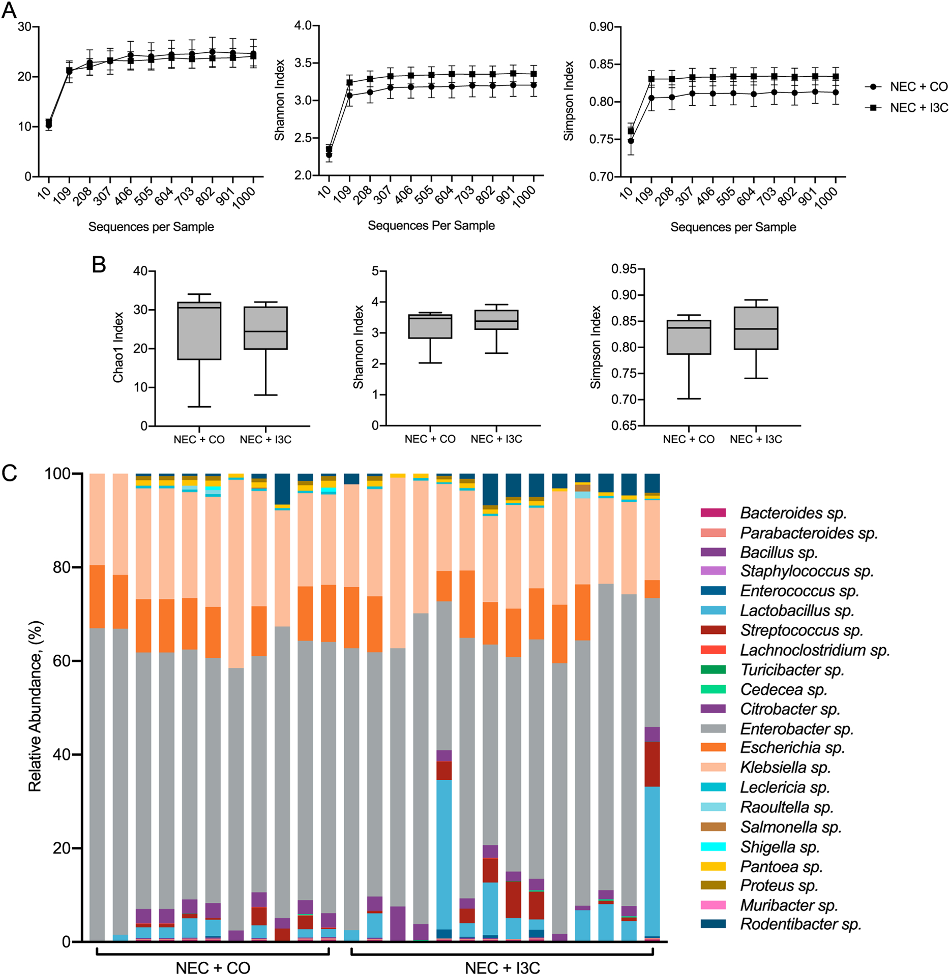
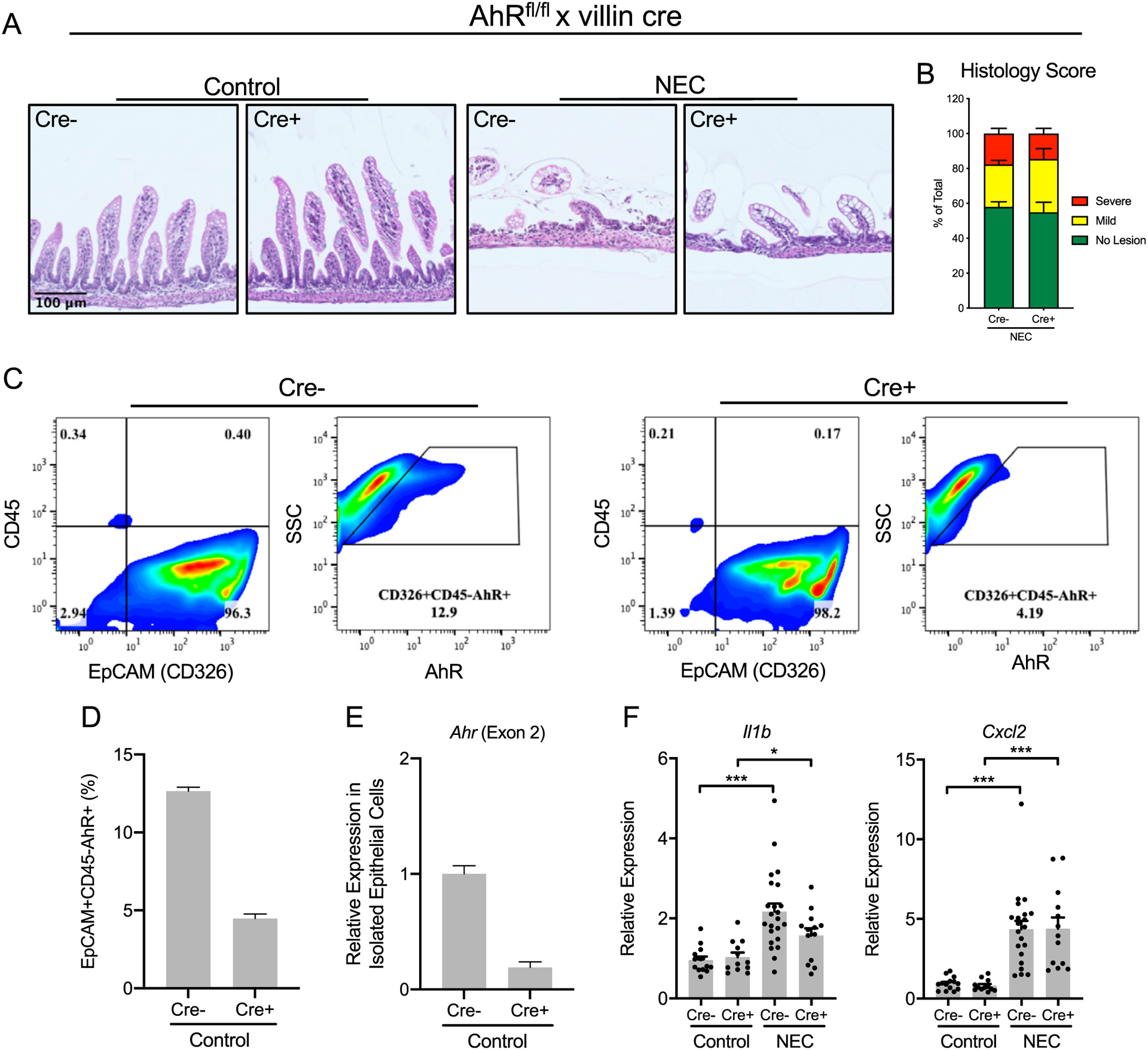
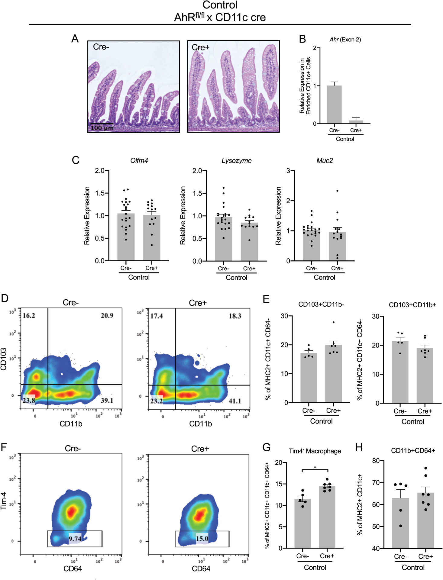
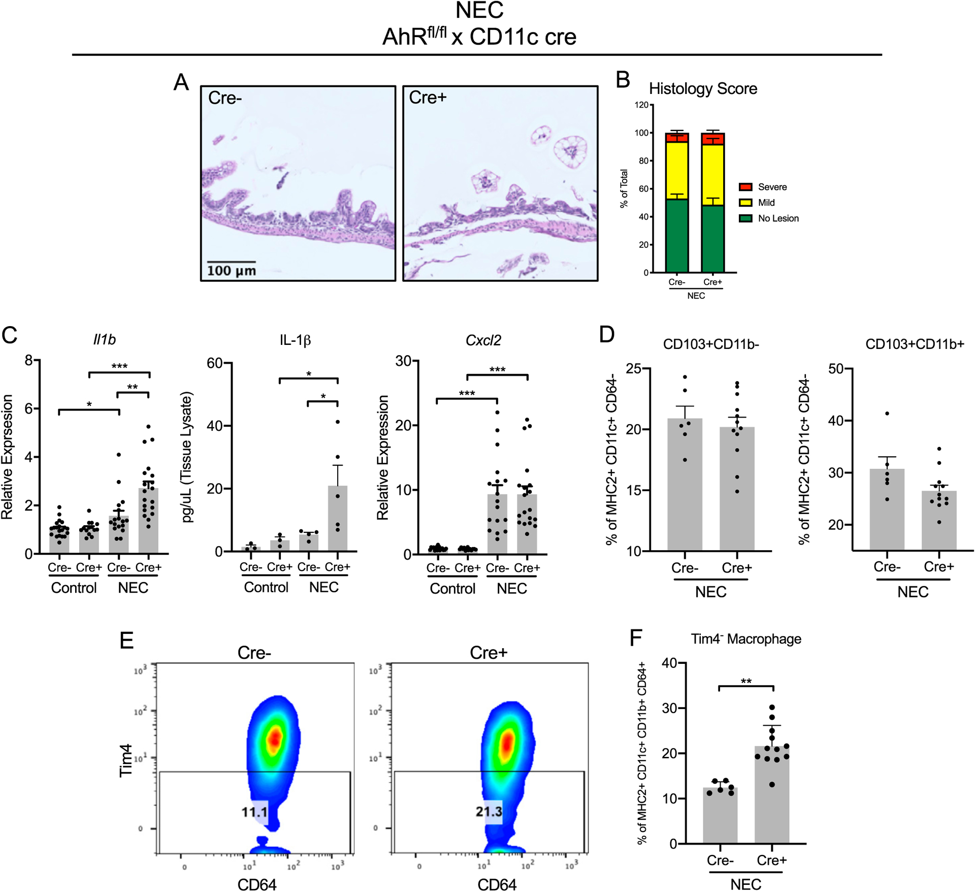
Necrotizing enterocolitis (NEC) causes significant morbidity and mortality in premature infants; therefore, the identification of therapeutic and preventative strategies against NEC remains a high priority. The ligand-dependent transcription factor aryl hydrocarbon receptor (AhR) is well known to contribute to the regulation of intestinal microbial communities and amelioration of intestinal inflammation. However, the role of AhR signaling in NEC is unclear. Experimental NEC was induced in 4-d-old wild-type mice or mice lacking AhR expression in the intestinal epithelial cells or AhR expression in CD11c+ cells (AhRΔCD11c) by subjecting animals to twice daily hypoxic stress and gavage feeding with formula supplemented with LPS and enteric bacteria. During NEC, compared with wild-type mice treated with vehicle, littermates treated with an AhR proligand, indole-3-carbinol, had reduced expression of Il1b and Marco, a scavenger receptor that mediates dendritic cell activation and the recognition and clearance of bacterial pathogens by macrophages. Furthermore, indole-3-carbinol treatment led to the downregulation of genes involved in cytokine and chemokine, as revealed by pathway enrichment analysis. AhR expression in the intestinal epithelial cells and their cre-negative mouse littermates were similarly susceptible to experimental NEC, whereas AhRΔCD11c mice with NEC exhibited heightened inflammatory responses compared with their cre-negative mouse littermates. In seeking to determine the mechanisms involved in this increased inflammatory response, we identified the Tim-4- monocyte-dependent subset of macrophages as increased in AhRΔCD11c mice compared with their cre-negative littermates. Taken together, these findings demonstrate the potential for AhR ligands as a novel immunotherapeutic approach to the management of this devastating disease.


Figures

FIGURE 1.
FIGURE 1.. I3C-mediated AhR signaling attenuates inflammation during experimental NEC.
AhR and CYP1A1 ontogeny are shown in (A) human fetal intestine for comparison with Ahr and Cyp1a1 ontogeny in (B) P1–49 mouse pups. (C) Treatment with AhR proligand I3C in control and experimental NEC pups produces a significant increase in Cyp1a1 expression, indicating activation of AhR signaling. (D) Representative images of formalin-fixed, paraffin-embedded murine terminal ileum tissue sections stained with H&E were used to quantify villi metrics and demonstrate I3C-mediated protection against intestinal barrier damage during NEC. (E) Blinded histology scoring quantitates the severity of intestinal lesions in the control and experimental groups with and without I3C treatment. (F) Terminal ileum expression of Il1b qRT-PCR (left) and protein concentration (right) is attenuated during NEC with I3C treatment. (G) Cxcl2, Lcn2, and Marco terminal ileal expression in wild-type control and I3C-fed pups with and without NEC. Data represent mean ± SEM from at least three independent experiments. *p < 0.05, **p < 0.005, ***p < 0.0001 by Mann–Whitney U test [(A)–(G) except (F), right, by unpaired t test]. CO, corn oil.
FIGURE 2.
FIGURE 2.. AhR activity during NEC induces a distinct profile of expressed proinflammatory response genes.
(A) Heat map representation of targeted genes in the experimental NEC groups with and without I3C exposure (fold change of at least 2-fold; p ≤ 0.05). (B) Volcano plot provides a comparison of gene expression in experimental NEC pups with I3C supplementation and demonstrates a profile of differentially expressed genes that are upregulated and downregulated during NEC. (C) Functionally enriched pathways provide a comparative analysis between the experimental NEC groups. Downregulation of chemokine signaling pathways and cytokine receptor interaction pathways occurred with AhR proligand supplementation during experimental NEC. CO, corn oil.
FIGURE 3.
FIGURE 3.. Enteral supplementation with I3C during NEC does not influence the gut microbiota.
The microbiota of fecal samples from murine NEC was evaluated using 16S ribosomal RNA-seq methods. (A and B) No significant difference in overall bacterial diversity was observed in the wild-type experimental NEC groups treated with I3C or the vehicle control, as shown by the Chao1, Shannon, and Simpson indices. (C) The relative abundance of selected bacteria in the experimental NEC groups is shown with no significant difference among the groups. CO, corn oil.
FIGURE 4.
FIGURE 4.. AhR signaling in IECs is dispensable in experimental NEC.
(A) Representative images of formalin-fixed, paraffin-embedded terminal ileum tissue sections from control and experimental pups were stained with H&E. (B) Tissue sections were used to quantify differences in histology scores in experimental pups with and without IEC-specific knockout without a significant difference in the proportion of lesions on NEC tissue sections. (C and D) Small intestine epithelial cells were isolated from AhRΔIEC pups (n = 2) and cre-negative littermates (n = 2) and stained with anti-EpCAM (anti-CD326) and anti-CD45, confirming positive isolation of epithelial cells and exclusion of lymphocytes. Flow cytometric analysis of isolated small IECs from cre-negative mice demonstrated a significantly increased percentage of AhR from EpCAM+CD45 cells when compared with AhRΔIEC mice, confirming epithelial cell-specific knockout of AhR. (E) qRT-PCR reveals significantly reduced mRNA expression of Ahr (Exon 2) in the isolated epithelial cells from AhRΔIEC pups, confirming AhR-specific deletion in the IECs. (F) AhRΔIEC pups exhibited a similar susceptibility to experimental NEC compared with their cre-negative littermates, without a significant difference in proinflammatory cytokine expression between the experimental NEC groups. *p < 0.05, ***p < 0.0001 by Mann-Whitney U test.
FIGURE 5.
FIGURE 5.. AhR signaling in CD11c+ immune cells is not induced during homeostasis.
(A) Representative images of formalin-fixed, paraffin-embedded terminal ileum tissue sections from mice with CD11c+ immune cell-specific knockout and cre-negative littermates during homeostasis were stained with H&E. (B) qRT-PCR analysis of enriched CD11c+ cells (continued) from the small intestine lamina propria from AhRΔCD11c pups (n = 2) and wild-type littermates (n = 2) reveals significantly reduced mRNA expression of Ahr (Exon 2) in AhRΔCD11c pups, confirming AhR-specific deletion in the CD11c+ immune cells. (C) Small intestine gene expression of IEC markers Olfm4, Lysozyme, and Muc2 were analyzed by qRT-PCR and demonstrated no change in the expression in the AhRΔCD11c pups or their cre-negative littermates. (D and E) Flow cytometric analysis demonstrated no significant change in the abundance of CD103+CD11b and CD103+CD11b+ DCs in cre-negative and AhRΔCD11c mice during steady state. (F and G) The Tim-4 monocyte–dependent subset of macrophages in the small intestine lamina propria were increased in AhRΔCD11c pups compared with cre-negative pups, whereas (H) there was no difference in CD11b+CD64+ macrophages in cre-negative mice and AhRΔCD11c mice during the homeostatic state. Data represent mean ± SEM from three independent experiments. *p < 0.05, by Mann-Whitney U test.
FIGURE 6.
FIGURE 6.. AhR signaling in CD11c+ immune cells impacts the severity of inflammation during NEC.
(A) Representative images of formalin-fixed, paraffin-embedded terminal ileum tissue sections stained with H&E are shown. (B) Tissue sections were used to quantify differences in histology scores in experimental pups with and without CD11c+ immune cell–specific knockout with no significant difference in the proportion of lesions on NEC tissue sections. (C) Relative expression of proinflammatory markers Il1b (left), quantitated by IL-1β protein concentration (middle), and Cxcl2 (right) in the small intestine were determined by qRT-PCR. (D) Flow cytometric analysis revealed no significant change in the abundance of CD103+CD11b or CD103+CD11b+ DCs in cre-negative and AhRΔCD11c mice during NEC. (E and F) Increased Tim-4 monocyte-dependent subset of macrophages in the small intestine lamina propria in AhRΔCD11c pups was observed during NEC. Data represent mean ± SEM from three independent experiments. *p < 0.05, **p < 0.005, ***p < 0.0001 by Mann-Whitney U test.

References

    1. Maloy KJ, and Powrie F. 2011. Intestinal homeostasis and its breakdown in inflammatory bowel disease. Nature 474: 298–306. - PubMed
    1. Neu J, and Walker WA. 2011. Necrotizing enterocolitis. N. Engl. J. Med 364: 255–264. - PMC - PubMed
    1. Lin PW, and Stoll BJ. 2006. Necrotising enterocolitis. Lancet 368: 1271–1283. - PubMed
    1. De Plaen IG 2013. Inflammatory signaling in necrotizing enterocolitis. Clin. Perinatol 40: 109–124. - PMC - PubMed
    1. Chng SH, Kundu P, Dominguez-Brauer C, Teo WL, Kawajiri K, Fujii-Kuriyama Y, Mak TW, and Pettersson S. 2016. Ablating the aryl hydrocarbon receptor (AhR) in CD11c+ cells perturbs intestinal epithelium development and intestinal immunity. Sci. Rep 6: 23820. - PMC - PubMed

Publication types

MeSH terms