. 2021 May;31(5):852-865.
doi: 10.1101/gr.266403.120. Epub 2021 Apr 27.

A novel fragmented mitochondrial genome in the protist pathogen Toxoplasma gondii and related tissue coccidia

Affiliations

A novel fragmented mitochondrial genome in the protist pathogen Toxoplasma gondii and related tissue coccidia

Sivaranjani Namasivayam et al. Genome Res. 2021 May.

Abstract

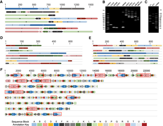
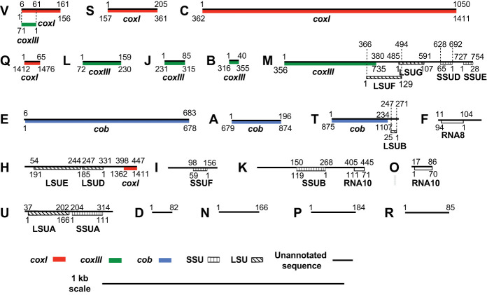
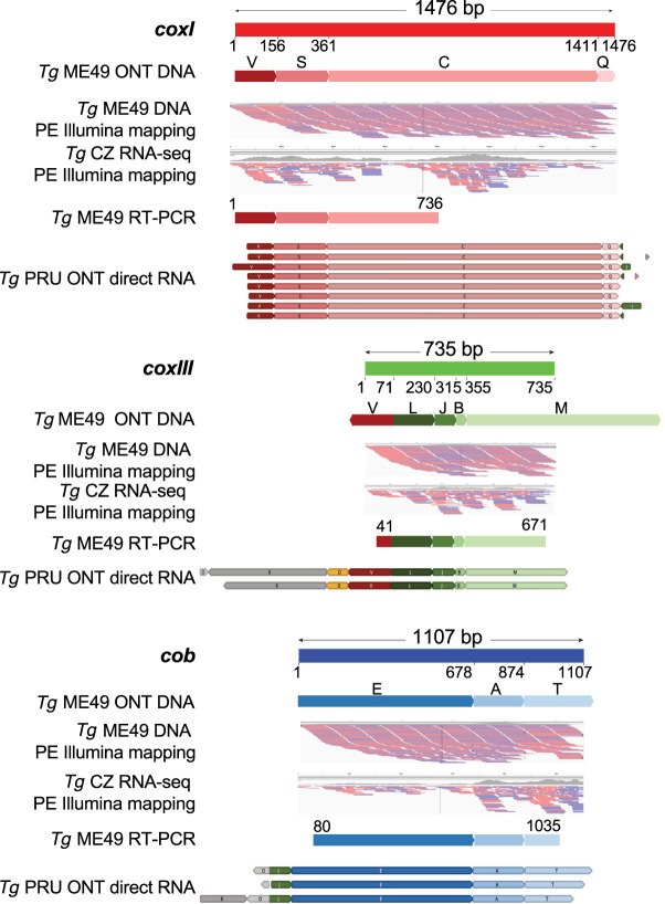
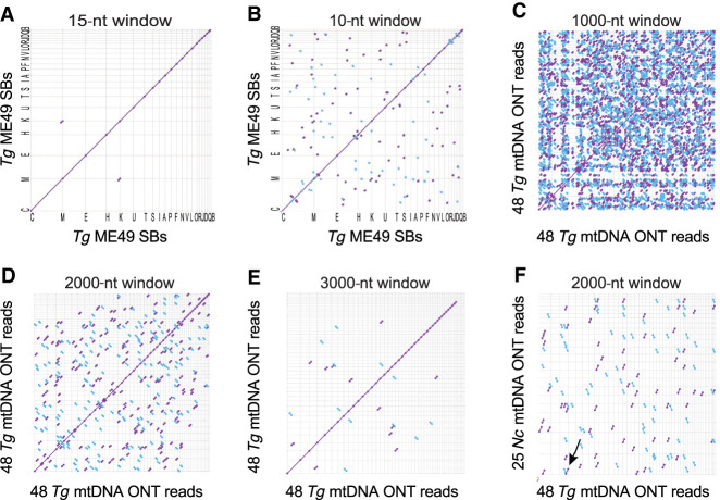
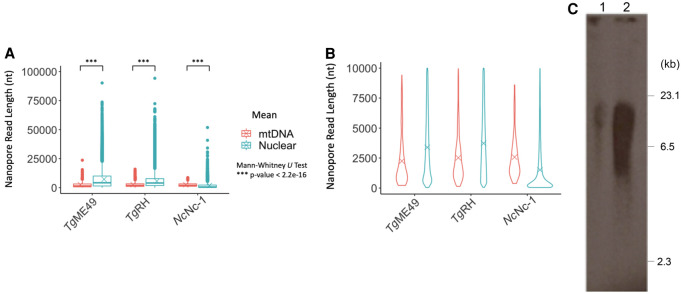
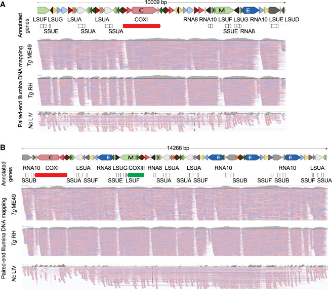
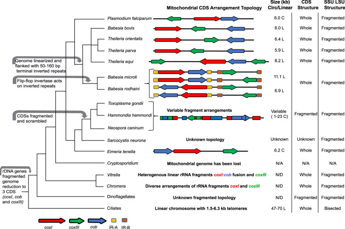
Mitochondrial genome content and structure vary widely across the eukaryotic tree of life, with protists displaying extreme examples. Apicomplexan and dinoflagellate protists have evolved highly reduced mitochondrial genome sequences, mtDNA, consisting of only three cytochrome genes and fragmented rRNA genes. Here, we report the independent evolution of fragmented cytochrome genes in Toxoplasma and related tissue coccidia and evolution of a novel genome architecture consisting minimally of 21 sequence blocks (SBs) totaling 5.9 kb that exist as nonrandom concatemers. Single-molecule Nanopore reads consisting entirely of SBs ranging from 0.1 to 23.6 kb reveal both whole and fragmented cytochrome genes. Full-length cytochrome transcripts including a divergent coxIII are detected. The topology of the mitochondrial genome remains an enigma. Analysis of a cob point mutation reveals that homoplasmy of SBs is maintained. Tissue coccidia are important pathogens of man and animals, and the mitochondrion represents an important therapeutic target. The mtDNA sequence has been elucidated, but a definitive genome architecture remains elusive.


Figures

Figure 1.
Figure 1.
Sequence evidence of fragmented mtDNA and full-length protein-encoding genes. (A, DF) Each row of colored blocks represents individual, annotated T. gondii (A) mtDNA-specific PCR amplicons; (D) EST reads; (E) Sanger genomic reads; or (F) Nanopore reads. Sections of reads containing identical sequence to other mtDNA reads are called “sequence blocks” (SBs) and are colored and labeled with a unique color and letter corresponding to the key, Table 1 and Figure 2. Shades of blue, red, and green represent different SBs found in cob, coxI, and coxIII, respectively. Orientation of a block is indicated by the point on each block. Red boxes in F indicate complete protein-encoding genes. SBs located on the ends of reads may be incomplete. (B) Genomic DNA from mitochondrial-enriched fractions assayed with different primer pairs (Supplemental Table S1) as indicated. Lanes 24 represent a small fragment of each cytochrome gene. (C) Single primers produce multiple amplicons. Scale (in bp) is as indicated in each panel. Additional reads are in Supplemental Figures S1–S4.
Figure 2.
Figure 2.
The 21 minimal T. gondii mtDNA sequence blocks. The DNA sequence is represented by a black line, drawn to scale and named with 21 alphabet characters, A to V (there is no “G”). The coordinates of an SB that encodes a cytochrome or rRNA gene fragment are indicated above the black line and the corresponding coordinates of the assembled gene or rRNA fragment are indicated below the gene fragment; the fragments are colored as defined in the key. Portions of sequence block V contribute to both coxI and coxIII but in different orientations.
Figure 3.
Figure 3.
Full-length protein-encoding genes and their expression in the T. gondii mt genome are supported. Segments of an ONT read that can encode a full-length cytochrome gene are annotated with their SB name and shown below the schematic of each gene. Numbers below each gene schematic represent nucleotide start/stop positions of a SB on the gene. All blocks are in the forward orientation except block V in coxIII. Only a portion of SBs V, M, and T is included in the CDS. MtDNA-specific PE-150 Illumina DNA (SRR9200762) and RNA-seq (SRR6493545) reads were independently mapped to each of the cytochrome gene sequences. Red and blue lines below each gene indicate mapped Illumina paired-ends. Both ends were required to map. RT-PCR products and ONT RNA reads with full-length CDSs are shown.
Figure 4.
Figure 4.
Dot plot comparisons of long T. gondii ME49 mtDNA SBs and ONT reads against themselves and N. caninum Nc-1. (A,B) T. gondii SBs (5909 nt) compared to themselves at differing window sizes; (CE) 48 mtDNA-specific T. gondii ONT reads (313,102 nt) (Supplemental Methods; Supplemental Fig. S4; Supplemental Data S3) compared to themselves at differing window sizes; (F) 25 N. caninum ONT mtDNA reads (141,161 nt) (Supplemental Methods; Supplemental Fig. S10; Supplemental Data S8) compared to the same 48 mtDNA-specific T. gondii ONT reads and visualized as a dot plot (see also Supplemental Fig. S11). Matches in the forward and reverse orientation are indicated as purple and blue lines, respectively. Sequences were sorted, from longest to shortest. Window size is indicated above each panel. The arrow in F points to the only match observed to be longer than 3000 bp. The names of the sequences located on each axis are located in the Supplemental Methods.
Figure 5.
Figure 5.
Size distribution of mtDNA versus nuclear ONT reads. (A) Three ONT runs are plotted and tested for statistical significance. Box plots cover the 25th–75th percentiles. See Supplemental Table S10 for mean and median values. (B) Violin plots of the same ONT reads <10 kb. (C) Southern of a CHEF gel electrophoresis of T. gondii total DNA (lane 1) and mitochondrial-enriched DNA (lane 2) probed with a 1012-bp section of the cob gene. DNA ladder is as indicated.
Figure 6.
Figure 6.
ONT and Illumina comparisons reveal sequence block order variability and decay with evolutionary time. (A,B) Two T. gondii ME49 ONT sequence reads were annotated with the gene sequences they contain. Reads and SBs are drawn to scale. The blocks are colored as shown in Table 1. “Annotated genes” track represents the annotation of the cytochrome genes and rRNA gene fragments on the ONT reads. The three “PE Illumina DNA mapping” tracks show PE read mapping of T. gondii ME49, TgRH88, and N. caninum (Nc) LIV mtDNA-specific reads. TgME49 and TgRH reads mapping required 100% nucleotide identity whereas 1% mismatch was allowed for mapping NcLIV reads. Reads were independently mapped to each of the ONT mtDNA reads and visualized using IGV. Red and blue lines below each read indicate the mapped Illumina PE reads. Coverage plots of the mapped reads are shown above each mapping panel in gray to indicate the depth of all reads and not just those shown.
Figure 7.
Figure 7.
Alveolate mtDNA evolution and characteristics. (Left) Cladogram of alveolate relationships with major mitochondrial genome events indicated with gray arrows. (Center) Schematic of mtDNA CDS (red, green, and blue arrows) or inverted repeats (gold and brown) indicated. Spacing is approximate with CDS lengths exaggerated for ease of viewing. Ribosomal RNA and other RNA genes or fragments thereof are not represented. (Right) mtDNA size and topology. (L) Linear, (C) concatemer, presumably from circular progenitors. Status of CDS sequences and status of Short (SSU) and Long Subunit (LSU) rRNA are indicated.

Similar articles

Cited by

References

    1. Alday PH, Bruzual I, Nilsen A, Pou S, Winter R, Ben Mamoun C, Riscoe MK, Doggett JS. 2017. Genetic evidence for cytochrome b Qi site inhibition by 4(1H)-quinolone-3-diarylethers and antimycin in Toxoplasma gondii. Antimicrob Agents Chemother 61: e01866-16. 10.1128/AAC.01866-16 - DOI - PMC - PubMed
    1. Bankevich A, Nurk S, Antipov D, Gurevich AA, Dvorkin M, Kulikov AS, Lesin VM, Nikolenko SI, Pham S, Prjibelski AD, et al. 2012. SPAdes: a new genome assembly algorithm and its applications to single-cell sequencing. J Comput Biol 19: 455–477. 10.1089/cmb.2012.0021 - DOI - PMC - PubMed
    1. Bedoya F, Medveczky MM, Lund TC, Perl A, Horvath J, Jett SD, Medveczky PG. 2009. Identification of mitochondrial genome concatemers in AIDS-associated lymphomas and lymphoid cell lines. Leuk Res 33: 1499–1504. 10.1016/j.leukres.2009.03.012 - DOI - PMC - PubMed
    1. Benson DA, Cavanaugh M, Clark K, Karsch-Mizrachi I, Ostell J, Pruitt KD, Sayers EW. 2018. Genbank. Nucleic Acids Res 46: D41–D47. 10.1093/nar/gkx1094 - DOI - PMC - PubMed
    1. Burger G, Lang BF. 2003. Parallels in genome evolution in mitochondria and bacterial symbionts. IUBMB Life 55: 205–212. 10.1080/1521654031000137380 - DOI - PubMed

Publication types

Substances

LinkOut - more resources