. 2021 Apr 27;11(1):9026.
doi: 10.1038/s41598-021-88506-y.

Colorimetric RT-LAMP SARS-CoV-2 diagnostic sensitivity relies on color interpretation and viral load

Affiliations

Colorimetric RT-LAMP SARS-CoV-2 diagnostic sensitivity relies on color interpretation and viral load

Mateus Nóbrega Aoki et al. Sci Rep. .

Abstract

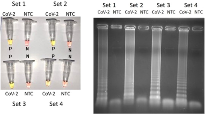
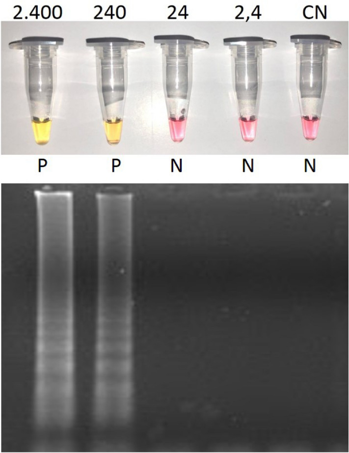
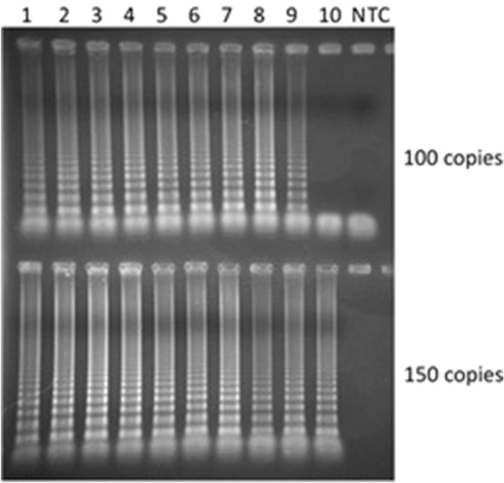
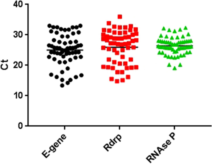
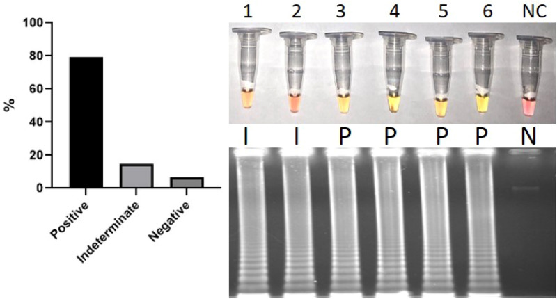
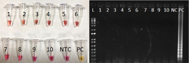
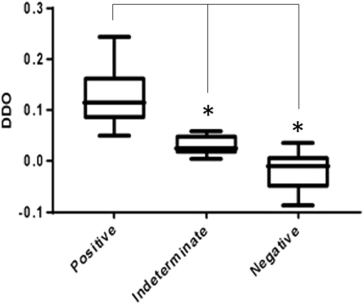
The use of RT-LAMP (reverse transcriptase-loop mediated isothermal amplification) has been considered as a promising point-of-care method to diagnose COVID-19. In this manuscript we show that the RT-LAMP reaction has a sensitivity of only 200 RNA virus copies, with a color change from pink to yellow occurring in 100% of the 62 clinical samples tested positive by RT-qPCR. We also demonstrated that this reaction is 100% specific for SARS-CoV-2 after testing 57 clinical samples infected with dozens of different respiratory viruses and 74 individuals without any viral infection. Although the majority of manuscripts recently published using this technique describe only the presence of two-color states (pink = negative and yellow = positive), we verified by naked-eye and absorbance measurements that there is an evident third color cluster (orange), in general related to positive samples with low viral loads, but which cannot be defined as positive or negative by the naked eye. Orange colors should be repeated or tested by RT-qPCR to avoid a false diagnostic. RT-LAMP is therefore very reliable for samples with a RT-qPCR Ct < 30 being as sensitive and specific as a RT-qPCR test. All reactions were performed in 30 min at 65 °C. The use of reaction time longer than 30 min is also not recommended since nonspecific amplifications may cause false positives.


Conflict of interest statement

The authors declare no competing interests.

Figures

Figure 1
Figure 1
Colorimetric RT-LAMP reaction for SARS-CoV-2 (2400 copies) using four primer sets: (A) yellow color (positive reaction indicated as P) and pink color (negative reaction indicated as N). (B) Amplification visualized in a 2% agarose gel stained with ethidium bromide, showing characteristic LAMP amplicon profiles in positive samples and no amplification in non-template controls (NTC). Set 1, Set 2, Sets 3 and 4.
Figure 2
Figure 2
The colorimetric and agarose gel detection of SARS-CoV-2 using RT-LAMP diluting a sample with 2400 copies of SARS-CoV-2 cultured in Vero cells (30 min, 65 °C, and primer set 3). Pink = negative (N); yellow = positive (P); CN = negative control.
Figure 3
Figure 3
Electrophoresis in 2% agarose gel stained with ethidium bromide and 10 samples of SARS-CoV-2 genome copies and amplified using the colorimetric RT-LAMP reaction at 65 °C for 30 min. All the samples containing 150 copies (100%) and 90% of the samples with 100 copies were amplified.
Figure 4
Figure 4
RT-qPCR samples Ct values for E-gene, RdRp gene and RNAse P (internal control).
Figure 5
Figure 5
Left panel: Diagnostic sensitivity of colorimetric RT-LAMP with 62 SARS-CoV-2 RT-qPCR positive samples: 79% positive (indicated as P), 14.5% indeterminate (I) and 6.5% negative (N). Right panel: Representative figure of SARS-CoV-2 clinical samples as indeterminate (1 and 2), positive (3 to 6) and a negative control (NC) using the colorimetric RT-LAMP method. The respective amplification product was also visualized in 2% agarose gel stained with ethidium bromide, showing the characteristic LAMP amplicons.
Figure 6
Figure 6
Representative Figure for non-SARS-CoV-2 samples present in Table 1. Samples: 1 and 2: FluA H1N1; 3 and 4: FluB Yamagata; 5 and 6: hCoV-229E; 7 and 8: hCoV-OC43; 9 and 10: hCoV-NL63; NTC: Non-template control; PC: Positive control; L: 1 kb Plus DNA ladder (ThermoFisher). As expected, only the PC changed color to yellow and was amplified in the agarose gel.
Figure 7
Figure 7
Delta DO for colorimetric RT-LAMP clinical samples measured by spectrophotometry clustering positive, indeterminate and negative results. Statistical significance (p < 0,05) was observed in ΔDO between three clusters.

Similar articles

Cited by

References

    1. Lai C-C, Shih T-P, Ko W-C, Tang H-J, Hsueh P-R. Severe acute respiratory syndrome coronavirus 2 (SARS-CoV-2) and coronavirus disease-2019 (COVID-19): The epidemic and the challenges. Int. J. Antimicrob. Agents. 2020;55:105924. doi: 10.1016/j.ijantimicag.2020.105924. - DOI - PMC - PubMed
    1. World Health Organization. Coronavirus Disease (COVID-19) Dashboard. https://covid19.who.int/ (2020).
    1. Fehr AR, Perlman S. Coronaviruses: an overview of their replication and pathogenesis. Methods Mol. Biol. 2015;1282:1–23. doi: 10.1007/978-1-4939-2438-7_1. - DOI - PMC - PubMed
    1. Montesinos I, et al. Evaluation of two automated and three rapid lateral flow immunoassays for the detection of anti-SARS-CoV-2 antibodies. J. Clin. Virol. 2020;128:104413. doi: 10.1016/j.jcv.2020.104413. - DOI - PMC - PubMed
    1. Chan JF-W, et al. Improved molecular diagnosis of COVID-19 by the novel, highly sensitive and specific COVID-19-RdRp/Hel real-time reverse transcription-pcr assay validated in vitro and with clinical specimens. J. Clin. Microbiol. 2020;58:e00310–e320. doi: 10.1128/JCM.00310-20. - DOI - PMC - PubMed

Publication types

Supplementary concepts