. 2022 Feb;43(2):483-493.
doi: 10.1038/s41401-021-00668-7. Epub 2021 Apr 27.

A multi-targeting drug design strategy for identifying potent anti-SARS-CoV-2 inhibitors

Affiliations

A multi-targeting drug design strategy for identifying potent anti-SARS-CoV-2 inhibitors

Peng-Xuan Ren et al. Acta Pharmacol Sin. 2022 Feb.

Abstract

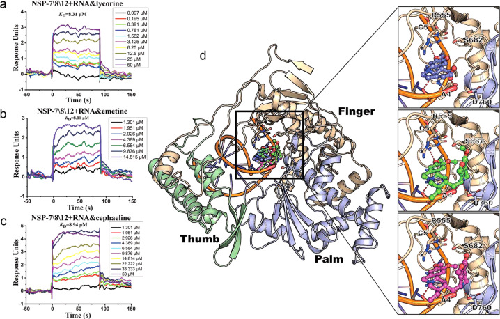
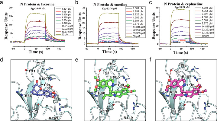
The COVID-19, caused by SARS-CoV-2, is threatening public health, and there is no effective treatment. In this study, we have implemented a multi-targeted anti-viral drug design strategy to discover highly potent SARS-CoV-2 inhibitors, which simultaneously act on the host ribosome, viral RNA as well as RNA-dependent RNA polymerases, and nucleocapsid protein of the virus, to impair viral translation, frameshifting, replication, and assembly. Driven by this strategy, three alkaloids, including lycorine, emetine, and cephaeline, were discovered to inhibit SARS-CoV-2 with EC50 values of low nanomolar levels potently. The findings in this work demonstrate the feasibility of this multi-targeting drug design strategy and provide a rationale for designing more potent anti-virus drugs.

Keywords: RdRp; SARS-CoV-2 inhibitors; Virus RNA; host ribosome.


Conflict of interest statement

The authors declare no competing interests.

Figures

Fig. 1
Fig. 1. Schematic illustration of the multi-targeting-based anti-SARS-CoV-2 inhibitor identification workflow designed by combining computational methods with experimental techniques.
The cartoon models represent the structures of four potential targets. The compound database is shown as a cylinder. The experimental techniques are shown as the pictures of instruments and equipments.
Fig. 2
Fig. 2. Computationally predicted molecular mechanism underlying the inhibition of viral translation of lycorine, emetine and cephaeline by targeting ribosome.
a Chemical structure of lycorine, emetine and cephaeline. b Predicted binding model of lycorine, emetine and cephaeline with human 80S ribosome. The 40S small subunit(palecyan) and the 60S large subunit(wheat) are shown in surface. Lycorine(blue), emetine(green) and cephaeline(magentas) bound at the active sites (E-site and PTC) are represented as stick-ball models. Three zoom-up models illustrate detailed molecular interactions between the ligands and the target. Red dash lines are hydrogen bonds or salt bridges formed between the ligand and targets.
Fig. 3
Fig. 3. Lycorine, emetine and cephaeline may attenuate SAR-CoV-2 propagation by binding with frame shift stimulation element (FSE).
Predicted binding modes of these ligands with FSE. The Cartoon model represents FSE (gray). Lycorine (blue), emetine (green) and cephaeline (magentas) bound at the binding sites are depicted as stick-ball models. The interaction ribonucleotides are depicted as sticks in the zoom-up models. Red dash lines are hydrogen bonds or salt bridges formed between the ligand and FSE. The secondary structure is shown at upper right.
Fig. 4
Fig. 4. Emetine and cephaeline may inhibit SARS-CoV-2 transcription by competitively blocking the binding of the RNA template-product duplex to RdRp.
a Representative dose-dependent response curves for lycorine, b emetine, c cephaeline binding with nsp-12 in solution obtained from SPR assay. The detailed kinetic data obtained from SPR is collected in Table S1. d The Cartoon model represents the catalytic part of nsp-12 protein including three domains of the finger (wheat), palm (lightblue) and thumb (palegreen). Lycorine (blue), emetine  (green) and cephaeline (magentas) bound at the active sites are represented as stick-ball models. The interaction residues are depicted as sticks in the zoom-up models. Red dash lines are hydrogen bonds or salt bridges formed between the ligand and proteins.
Fig. 5
Fig. 5. Lycorine, emetine and cephaeline may inhibit SARS-CoV-2 transcription by interfering with the function of RNA on its growth site of RdRp.
a Representative dose-dependent response curves for lycorine, b emetine, c cephaeline binding with nsp-7\8\12 and RNA complex in solution obtained from SPR assay. d Predicted binding modes of these ligands with the complex of RdRp and RNA. The Cartoon model represents the catalytic part of nsp-12 protein including three domains of the finger (wheat), palm (lightblue) and thumb (palegreen) domains. Lycorine (blue), emetine (green) and cephaeline (magentas) bound at the active sites are depicted as stick-ball models. RNA is shown as yellow cartoon model. The interaction residues are depicted as sticks in the zoom-up models. Red dash lines are hydrogen bonds or salt bridges formed between the ligand and proteins.
Fig. 6
Fig. 6. Lycorine, emetine and cephaeline may inhibit the assembly of SARS-CoV-2 virions.
a Representative response curve for lycorine, b emetine, c cephaeline binding with N protein in solution, resulting from SPR assay. The detailed kinetic data obtained from SPR are collected in Table S2. d–f Predicted binding modes of these ligands with N protein. The cartoon model represents N protein (palecyan). Lycorine (blue), emetine (green) and cephaeline (magentas) bound at the active sites are depicted as stick-ball models. The interaction residues are depicted as sticks. Red dash lines are hydrogen bonds or salt bridges formed between the ligand and proteins.
Fig. 7
Fig. 7. In vitro inhibitory activity profiles of compounds against SARS-CoV-2.
Vero E6 cells infected with SARS-CoV-2 at an MOI of 0.01 were treated with different concentrations of compounds. a Quantitative RT-PCR assays were performed to measure the viral copy number in cellular supernatant. The y axis indicates percentage inhibition of virus relative to sample treated with DMSO (vehicle); b Cell viability assay in Vero E6 cells. The y axis represents the percentage of cell viability relative to the sample treated with DMSO (vehicle). Data are shown as mean ± SD, n = 3.

Similar articles

Cited by

References

    1. Zhu N, et al. A novel coronavirus from patients with pneumonia in China, 2019. N Engl J Med. 2020;382:727–33. - PMC - PubMed
    1. Holshue ML, et al. First case of 2019 novel coronavirus in the United States. N Engl J Med. 2020;382:929–36. - PMC - PubMed
    1. Lillie PJ, et al. Novel coronavirus disease (Covid-19): The first two patients in the UK with person to person transmission. J Infect. 2020;80:578–606. - PMC - PubMed
    1. Dong E, Du H, Gardner L. An interactive web-based dashboard to track COVID-19 in real time. Lancet Infect Dis. 2020;20:533–4. - PMC - PubMed
    1. Zhou P, et al. A pneumonia outbreak associated with a new coronavirus of probable bat origin. Nature. 2020;579:270–3. - PMC - PubMed

Substances