. 2021 Apr 20:16:2949-2963.
doi: 10.2147/IJN.S304515. eCollection 2021.

Extracellular Vesicles from Akkermansia muciniphila Elicit Antitumor Immunity Against Prostate Cancer via Modulation of CD8+ T Cells and Macrophages

Affiliations

Extracellular Vesicles from Akkermansia muciniphila Elicit Antitumor Immunity Against Prostate Cancer via Modulation of CD8+ T Cells and Macrophages

Zhong-Wei Luo et al. Int J Nanomedicine. .

Abstract

Purpose: Prostate cancer (PCa) is one of the most common malignancies in males. Despite the success of immunotherapy in many malignant cancers, strategies are still needed to improve therapeutic efficacy in PCa. This study aimed to investigate the effects of Akkermansia muciniphila-derived extracellular vesicles (Akk-EVs) on PCa and elucidate the underlying immune-related mechanism.

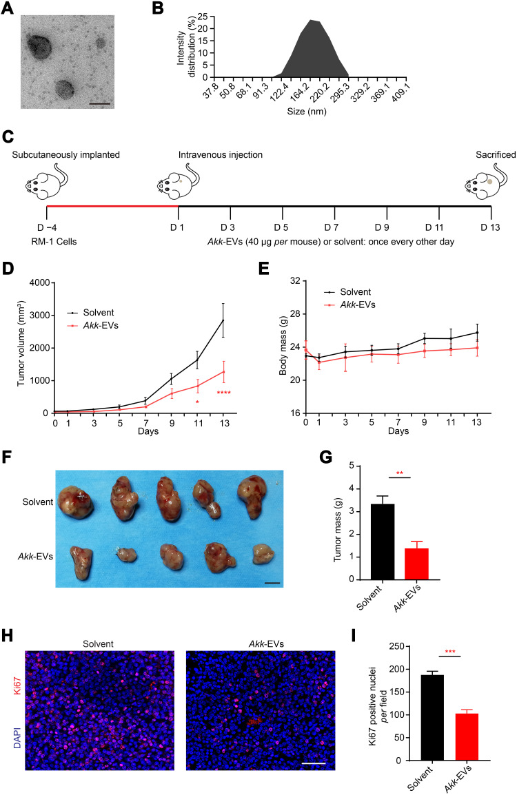
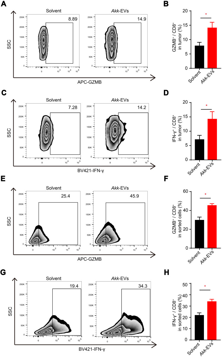
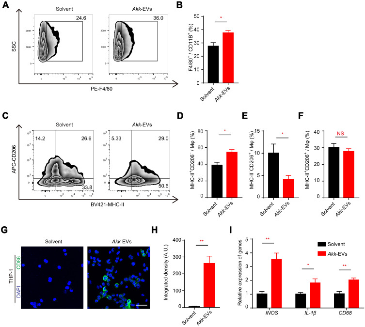
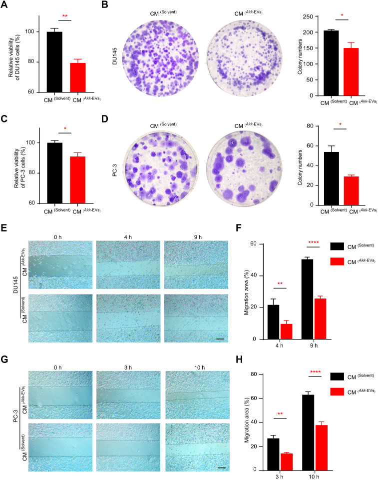
Methods: Akk-EVs were isolated by ultracentrifugation and intravenously injected to treat syngeneic PCa-bearing immune-competent mice. Immunophenotypic changes in immune cells, such as cytotoxic T lymphocytes and macrophages, were measured via flow cytometry analysis. Histological examination was used to detect morphological changes in major organs after Akk-EVs treatments. In vitro, flow cytometry was performed to confirm the effects of Akk-EVs on the activation of CD8+ T cells. Quantitative PCR and immunofluorescence staining were carried out to test the impact of Akk-EVs on macrophage polarization. Cell counting kit-8 (CCK-8) analysis, colony formation assays, and scratch wound healing assays were conducted to assess the effects of Akk-EVs-treated macrophages on the proliferation and invasion of PCa cells. CCK-8 assays also confirmed the impact of Akk-EVs on the viability of normal cells.

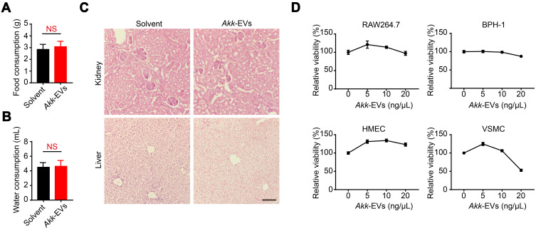
Results: Intravenous injection of Akk-EVs in immune-competent mice reduced the tumor burden of PCa without inducing obvious toxicity in normal tissues. This treatment elevated the proportion of granzyme B-positive (GZMB+) and interferon γ-positive (IFN-γ+) lymphocytes in CD8+ T cells and caused macrophage recruitment, with increased tumor-killing M1 macrophages and decreased immunosuppressive M2 macrophages. In vitro, Akk-EVs increased the number of GZMB+CD8+ and IFN-γ+CD8+ T cells and M1-like macrophages. In addition, conditioned medium from Akk-EVs-treated macrophages suppressed the proliferation and invasion of prostate cells. Furthermore, the effective dose of Akk-EVs was well-tolerated in normal cells.

Conclusion: Our study revealed the promising prospects of Akk-EVs as an efficient and biocompatible immunotherapeutic agent for PCa treatment.

Keywords: Akkermansia muciniphila; cytotoxic T lymphocytes; extracellular vesicles; immunotherapy; macrophages; prostate cancer.


Conflict of interest statement

The authors report no conflicts of interest in this work.

Figures

Figure 1
Figure 1
Akk-EVs reduced tumor burden in a PCa mouse model. (A) Representative TEM image of Akk-EVs. Scale bar: 200 nm. (B) The particle size distribution of Akk-EVs detected by DLS. (C) Schematic diagram of the treatment schedule for subcutaneous RM-1 tumor-bearing C57BL/6 mice. (D) Tumor volumes of mice in different treatment groups are shown at the indicated times during the observation period of 13 days. n = 5 per group. (E) Body mass of mice were recorded during the whole experiment. n = 5 per group. (F and G) Digital photographs (F) and the mass (G) of tumor tissues isolated from the RM-1 tumor-bearing mice on the 13th day after different treatments. Scale bar: 1 cm. n = 5 per group. (H and I) Representative images (H) and quantification (I) of Ki67 immunofluorescence staining of tumor sections. Scale bar: 50 μm. n = 3 per group. Data are shown as mean ± SEM. *P < 0.05, **P < 0.01, ***P < 0.001, ****P < 0.0001.
Figure 2
Figure 2
Akk-EVs elevated the proportion of GZMB+ and IFN-γ+ lymphocytes in CD8+ T cells in vivo and in vitro. (AD) Flow cytometry analysis of the number of GZMB and IFN-γ of CD8+ T cells infiltrated in PCa tumor tissues of solvent or Akk-EVs-treated mice. Representative plots (A and C) and quantitative analysis (B and D) were shown. n = 5 per group. (EH) Flow cytometry analysis of GZMB and IFN-γ expression of CD8+ T cells in vitro. Representative plots (E and G) and quantitative analysis (F and H). n = 5 per group. Data are shown as mean ± SEM. *P < 0.05.
Figure 3
Figure 3
Akk-EVs recruited macrophages and skewed them to an M1-like phenotype. (AF) Flow cytometry analysis of tumor infiltrated macrophages of solvent- or Akk-EVs-treated mice. Macrophages (Mφ) were identified as CD11b+F4/80+ cells. Representative plots (A) and quantitative analysis (B) of the proportion of F4/80+ cells in CD11b+ cells were shown. M1-like and M2-like Mφ were identified as MHC-II+CD206 and MHC-IICD206+, respectively. Representative plots (C) showed the gating strategy to define different Mφ phenotypes, and quantitative analysis of different subpopulations was also shown (DF). n = 5 per group. (G and H) Representative images (G) and quantification (H) of CD86-stained PMA-pretreated THP-1 after solvent or Akk-EVs treatment for 48h. Scale bar: 50 μm. (I) qRT-PCR analysis of the expression levels of INOS, CD68, and IL-1β in PMA-pretreated human THP-1 cells receiving different treatments for 24 h. n = 3 per group. Data are shown as mean ± SEM. *P < 0.05, **P < 0.01. NS, not significant.
Figure 4
Figure 4
Akk-EVs-treated macrophages suppressed the proliferation and invasion of prostate cells. (A and C) CCK-8 analysis of the viability of human PCa cell line DU145 (A) and PC-3 (C) cells receiving different CM treatments for 24 h. n = 4 per group. (B and D) Representative images and quantitative analysis of the crystal violet-stained colonies formed by DU145 (B) and PC-3 (D) cells receiving different treatments for 14 days. n = 3 per group. (EH) Representative images of scratch wound healing assay in DU145 (E) and PC-3 (G) cells after different CM treatments at indicated time points and corresponding quantification of the migration rates in DU145 (F) and PC-3 (H). Scale bar: 200 μm. n = 4 per group. Data are shown as mean ± SEM. *P < 0.05, **P < 0.01, ****P < 0.0001.
Figure 5
Figure 5
Akk-EVs were tolerated in vivo and in vitro. (A and B) Average daily food (A) and water (B) consumption of mice were shown. n = 5 per group. (C) Representative H&E staining images of kidney and liver from subcutaneous RM-1 bearing mice treated with solvent or Akk-EVs for 13 days. Scale bar: 100 μm. n = 3 per group. (D) CCK-8 analysis of the viability of a panel of normal cell lines (RAW264.7, BPH-1, HMECs, and VSMC) receiving different doses of Akk-EVs for 24 h. n = 4 per group. Data are shown as mean ± SEM. NS, not significant.

Similar articles

Cited by

References

    1. Siegel RL, Miller KD, Jemal A. Cancer statistics, 2020. CA Cancer J Clin. 2020;70(1):7–30. doi:10.3322/caac.21590 - DOI - PubMed
    1. Culp MB, Soerjomataram I, Efstathiou JA, Bray F, Jemal A. Recent global patterns in prostate cancer incidence and mortality rates. Eur Urol. 2020;77(1):38–52. doi:10.1016/j.eururo.2019.08.005 - DOI - PubMed
    1. Nuhn P, De Bono JS, Fizazi K, et al. Update on systemic prostate cancer therapies: management of metastatic castration-resistant prostate cancer in the era of precision oncology. Eur Urol. 2019;75(1):88–99. doi:10.1016/j.eururo.2018.03.028 - DOI - PubMed
    1. Ku SY, Gleave ME, Beltran H. Towards precision oncology in advanced prostate cancer. Nat Rev Urol. 2019;16(11):645–654. doi:10.1038/s41585-019-0237-8 - DOI - PMC - PubMed
    1. Powers E, Karachaliou GS, Kao C, et al. Novel therapies are changing treatment paradigms in metastatic prostate cancer. J Hematol Oncol. 2020;13(1):144. doi:10.1186/s13045-020-00978-z - DOI - PMC - PubMed

MeSH terms

Substances

Supplementary concepts