. 2021 Apr 19:13:3429-3442.
doi: 10.2147/CMAR.S290966. eCollection 2021.

Matrine Regulates Proliferation, Apoptosis, Cell Cycle, Migration, and Invasion of Non-Small Cell Lung Cancer Cells Through the circFUT8/miR-944/YES1 Axis

Affiliations

Matrine Regulates Proliferation, Apoptosis, Cell Cycle, Migration, and Invasion of Non-Small Cell Lung Cancer Cells Through the circFUT8/miR-944/YES1 Axis

Hailing Zhu et al. Cancer Manag Res. .

Retraction in

Abstract

Background: Non-small cell lung carcinoma (NSCLC) is the major histological subtype of cancer cases. In the present study, we investigated the association between Matrine, an active component of Chinese medicine, and circFUT8 in NSCLC cells.

Methods: The proliferation ability of NSCLC cells was assessed by MTT and colony-forming assays. Flow cytometry assay was performed to show the apoptosis and cell cycle distribution in NSCLC cells. The protein expression levels of Bcl-2, Cleaved Caspase-3 (C-Caspase3), and YES proto-oncogene 1 (YES1) were measured by Western blot assay. Migration and invasion of NSCLC cells were determined by transwell assay. The expression levels of circFUT8, miR-944 and YES1 were quantified by real-time quantitative polymerase chain reaction (RT-qPCR) assay. The interaction relationship between miR-944 and circFUT8 or YES1 was confirmed by dual-luciferase reporter assay. The anti-tumor role of Matrine in vivo was explored by a xenograft experiment.

Results: Matrine functioned as a carcinoma inhibitor by repressing proliferation, cell cycle process, migration, and invasion while inducing apoptosis in NSCLC cells. Importantly, overexpression of circFUT8 counteracted Matrine-induced effects on NSCLC cells. MiR-944, interacted with YES1, was a target of circFUT8. Under Matrine condition, overexpression of circFUT8 increased proliferation, migration, and invasion while inhibited apoptosis, which was abolished by the upregulation of miR-944. Whereas the silencing of YES1 counteracted miR-944 inhibitor-induced effects on NSCLC cells. Eventually, we also confirmed that Matrine impeded NSCLC tumor growth in vivo.

Conclusion: Matrine regulated proliferation, apoptosis, cell cycle, migration, and invasion of NSCLC cells through the circFUT8/miR-944/YES1 axis, which provided novel information for Matrine in NSCLC.

Keywords: Matrine; NSCLC; YES1; circFUT8; miR-944.


Conflict of interest statement

The authors declare that they have no financial or non-financial conflicts of interest for this work.

Figures

Figure 1
Figure 1
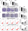
The effects of Matrine on proliferation, apoptosis, cell cycle, migration, and invasion in NSCLC cells. (AL) H522 and H1975 cells were treated with Matrine with different concentrations (0, 0.5, 1, and 2 mg/mL) for 24 h. (AD) The proliferation ability of H522 and H1975 cells were measured by MTT and colony-forming assays. (E and F) The flow cytometry assay was performed to monitor apoptotic cells. (G and H) The protein expression levels of Bcl-2 and C-Caspase3 were detected by Western blot assay. (I and J) The cell cycle distribution was shown by flow cytometry assay. (K and L) The migration and invasion of H522 and H1975 cells were determined by transwell assay. *P < 0.05.
Figure 1
Figure 1
continued.
Figure 2
Figure 2
The expression level of circFUT8 in NSCLC tissues and cells. (A) The expression level of circFUT8 was shown from GEO accession. (B) CircFUT8 level was measured by RT-qPCR assay in NSCLC cells and control group. (C and D) RT-qPCR assay was conducted to show expression of circFUT8 and linearFUT8 in H522 and H1975 cells after treatment with RNase R. (E and F) The abundant of circFUT8 in the cytoplasm and nucleus was quantified by RT-qPCR assay. (G and H) After being treated by Matrine, the level of circFUT8 was shown by RT-qPCR assay in H522 and H1975 cells. *P < 0.05.
Figure 3
Figure 3
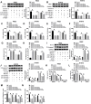
Effects of circFUT8 overexpression on proliferation, apoptosis, cell cycle, migration, and invasion in NSCLC cells treated with Matrine. (AN) H522 and H1975 cells were divided into four groups: Control, Matrine, Matrine+vector, and Matrine+circFUT8. (A and B) The expression level of circFUT8 was examined by RT-qPCR assay after transfection. (CF) MTT and colony-forming assays were used to measuring cell proliferation. (G and H) The apoptosis rate of H522 and H1975 cells was shown by flow cytometry assay. (I and J) The Western blot assay was performed to assess the expression of Bcl-2 and C-Caspase3 in H522 and H1975 cells. (K and L) The cell cycle distribution was shown by flow cytometry assay. (M and N) The transwell assay was conducted in H522 and H1975 cells post-transfection. *P < 0.05.
Figure 4
Figure 4
MiR-944 was a direct target of circFUT8 in NSCLC cells. (A) Binding regions between miR-944 and circFUT8 were shown. (B and C) The relative luciferase activity was analyzed in H522 and H1975 cells co-transfected with reports and miR-944 mimic or miR-NC. (D and E) RT-qPCR assay was conducted to evaluate miR-944 level in H522 and H1975 cells transfected with vector or circFUT8. (F and G) The expression level of miR-944 was assessed by RT-qPCR assay in H522 and H1975 cells exposed to Matrine with different concentrations (0, 0.5, 1, and 2 mg/mL) for 24 h. *P < 0.05.
Figure 5
Figure 5
Matrine regulated proliferation, apoptosis, cell cycle, migration, and invasion of NSCLC cells through circFUT8/miR-944 axis. (AN) H522 and H1975 cells were divided into six groups: Control, Matrine, Matrine+vector, Matrine+circFUT8, Matrine+circFUT8+miR-NC, and Matrine+circFUT8+miR-944. (A and B) The expression level of miR-944 was detected by RT-qPCR assay. (CF) The proliferation capability of H522 and H1975 cells was analyzed by MTT and colony-forming assays. (G and H) The flow cytometry assay was used to show the apoptosis rate of H522 and H1975 cells. (I and J) The expression levels of Bcl-2 and C-Caspase3 were calculated with Western blot assay. (K and L) The cell cycle distribution was shown by flow cytometry assay. (M and N) The transwell assay was performed to assess migration and invasion of H522 and H1975 cells post-transfection. *P < 0.05.
Figure 6
Figure 6
CircFUT8/miR-944 axis regulated YES1 expression in NSCLC cells. (A) MiR-944 had the binding region in 3ʹUTR of YES1 mRNA. (B and C) The luciferase activities of the wild-type YES1 3ʹ UTR WT and mutant YES1 3ʹ UTR MUT were measured. (D and E) Western blot assay was performed to assess YES1 level in H522 and H1975 cells transfected with anti-miR-NC or anti-miR-944. (F and G) After treatment with Matrine with different concentrations (0, 0.5, 1, and 2 mg/mL) for 24 h, the expression of YES1 was shown by Western blot assay. (H and I) The protein expression level of YES1 was determined by Western blot assay in H522 and H1975 cells transfected with vector, circFUT8, circFUT8+miR-NC, or circFUT8+miR-944. *P < 0.05.
Figure 7
Figure 7
MiR-944/YES1 regulated proliferation, apoptosis, cell cycle, migration, and invasion of NSCLC cells exposed to Matrine. (AN) H522 and H1975 cells were divided into six groups: Control, Matrine, Matrine+anti-miR-NC, Matrine+anti-miR-944, Matrine+anti-miR-944+si-NC, or Matrine+anti-miR-944+si-YES1. (A and B) The protein expression level of YES1 was evaluated by Western blot assay. (CF) MTT and colony-forming assays were conducted to assess cell proliferation. (G and H) The apoptosis rate was determined by flow cytometry assay. (I and J) The expression levels of Bcl-2 and C-Caspase3 were quantified with Western blot assay. (K and L) The cell cycle distribution was displayed by flow cytometry assay. (M and N) The migration and invasion were measured by transwell assay in H522 and H1975 cells. *P < 0.05.
Figure 8
Figure 8
Matrine repressed NSCLC tumor growth in vivo. (A and B) The volume and weight of xenograft tumors were shown. (C and D) The expression levels of circFUT8 and miR-944 were estimated with RT-qPCR assay. (E) Western blot assay was used to show the expression level of YES1 in tumor tissues, with GAPDH as control. (F) Immunohistochemistry for ki-67 and C-Caspase3 was performed in tumor tissues. *P < 0.05.

Similar articles

Cited by

References

    1. Bray F, Ferlay J, Soerjomataram I, Siegel RL, Torre LA, Jemal A. Global cancer statistics 2018: GLOBOCAN estimates of incidence and mortality worldwide for 36 cancers in 185 countries. CA Cancer J Clin. 2018;68(6):394–424. doi:10.3322/caac.21492 - DOI - PubMed
    1. Hirsch FR, Scagliotti GV, Mulshine JL, et al. Lung cancer: current therapies and new targeted treatments. Lancet. 2017;389(10066):299–311. doi:10.1016/S0140-6736(16)30958-8 - DOI - PubMed
    1. Xu N, Chen S, Liu Y, et al. Profiles and bioinformatics analysis of differentially expressed circrnas in taxol-resistant non-small cell lung cancer cells. Cell Physiol Biochem. 2018;48(5):2046–2060. doi:10.1159/000492543 - DOI - PubMed
    1. Li J, Yang J, Zhou P, et al. Circular RNAs in cancer: novel insights into origins, properties, functions and implications. Am J Cancer Res. 2015;5(2):472. - PMC - PubMed
    1. Yuan W, Peng S, Wang J, et al. Identification and characterization of circRNAs as competing endogenous RNAs for miRNA-mRNA in colorectal cancer. PeerJ. 2019;7:e7602. doi:10.7717/peerj.7602 - DOI - PMC - PubMed

Publication types