

Synthesis of intracellular and extracellular gold nanoparticles with a green machine and its antifungal activity
- PMID: 33907501
- PMCID: PMC8068771
- DOI: 10.3906/biy-2010-64
Synthesis of intracellular and extracellular gold nanoparticles with a green machine and its antifungal activity
Abstract
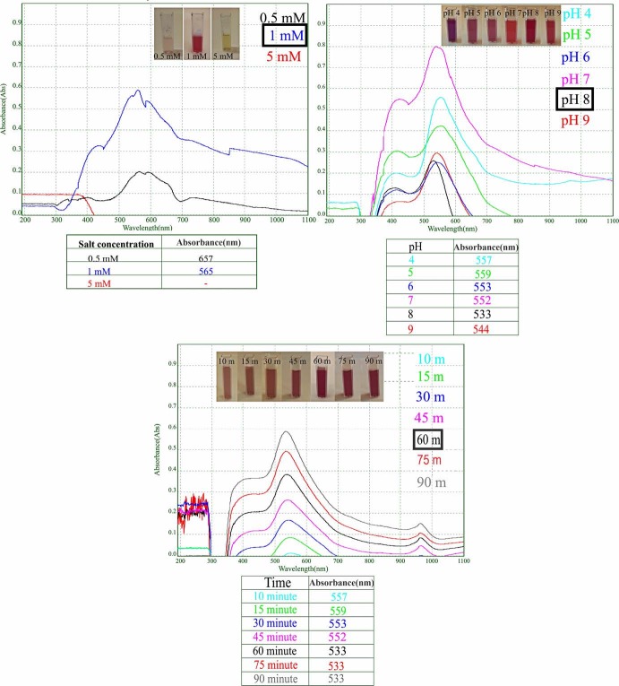
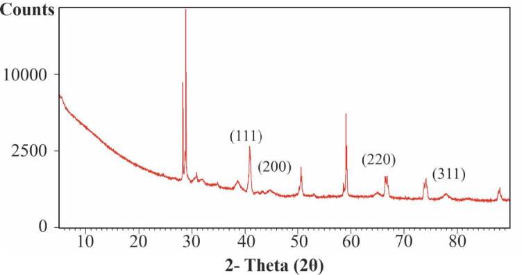
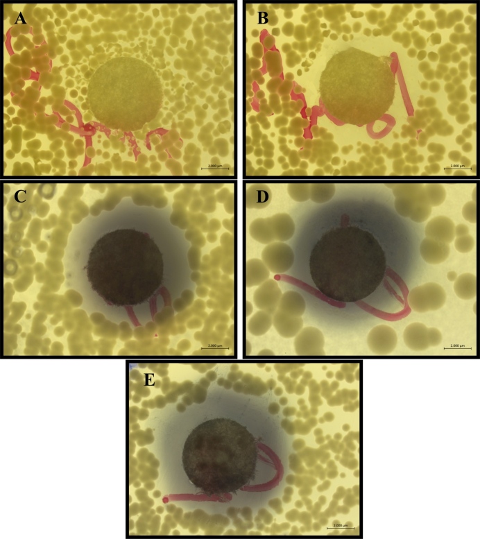
Green synthesis method is being increasingly used in the development of safe, stable, and eco-friendly nanostructures with biological resources. In this study, extracellular and intracellular synthesis of gold nanoparticles (AuNPs) was carried out using green algae Chlorella sorokiniana Shihira & R.W. Fresh algae were isolated and identified from Musaözü Pond located in the province of Eskişehir and then extraction process were performed. Optimization studies were studied using pH value, metal salt concentration, and time parameters for extracellular synthesis and using only time parameter for intrasellular synthesis. Since more controlled and optimum conditions can be achieved in the production of AuNPs by extracellular synthesis, these nanoparticles (NPs) were used for characterization and antifungal activity studies. Optical, physical, and chemical properties of synthesized NPs were characterized by UV visible spectrophotometer (UV-Vis), dynamic light scattering (DLS), Zetasizer, X-Ray diffraction (XRD), Fourier transform ınfrared spectroscopy (FTIR), field emission scanning electron microscope (FE-SEM), ınductively coupled plasma mass spectrometer (ICP-MS) and transmission electron microscope (TEM) analysis. The optimum conditions for AuNPs synthesis were determined as 1 mM for HauCl4 concentration, 6 for pH value, and 60th min for time. AuNPs obtained from extracellular synthesis from C. sorokiniana extract are 5-15 nm in size and spherical shape. TEM images of extracellular synthesis show noticeable cell wall and membrane damages, cytoplasma dissolutions, and irregularities. AuNPs obtained by intracellular synthesis are in 20-40 nm size and localized in the cell wall and cytoplasm. These NPs exhibited significant antifungal activity against C. tropicalis, C. glabrata, and C. albicans isolates. AuNPs obtained by algae-mediated green synthesis have a significant potential for medical and industrial use, and this eco-friendly synthesis method can be easily scaled for future studies.
Keywords: Chlorella; antifungal; gold nanoparticles; green synthesis; transmission electron microscope (TEM).
Copyright © 2021 The Author(s).
Conflict of interest statement
CONFLICT OF INTEREST: The authors state no conflict of interest.
Figures


References
-
- Abdel-Raouf N Al-Enazi NM Ibraheem IB Green biosynthesis of gold nanoparticles using Galaxaura elongata and characterization of their antibacterial activity. Arabian Journal of Chemistry. 2017;10:S3029–S3039.
-
- Aboelfetoh EF El-Shenody RA Ghobara MM Eco-friendly synthesis of silver nanoparticles using green algae (Caulerpa serrulata): reaction optimization, catalytic and antibacterial activities. Environmental Monitoring and Assessment. 2017;189:349–349. - PubMed
-
- Ahmed S Ikram S Biosynthesis of gold nanoparticles: a green approach. Journal of Photochemistry and Photobiology B: Biology. 2016;161:141–153. - PubMed
-
- Andersen RA Kawachi M Microalgae isolation techniques. Algal Culturing Techniques. 2005;83
-
- Annamalai J Nallamuthu T Characterization of biosynthesized gold nanoparticles from aqueous extract of Chlorella vulgaris and their anti-pathogenic properties. Applied Nanoscience. 2015;5:603–607.
LinkOut - more resources
Full Text Sources
Other Literature Sources