Review
. 2021 Jul 15:893:115289.
doi: 10.1016/j.jelechem.2021.115289. Epub 2021 Apr 23.

Emerging materials for the electrochemical detection of COVID-19

Affiliations
Review

Emerging materials for the electrochemical detection of COVID-19

G Balkourani et al. J Electroanal Chem (Lausanne). .

Abstract

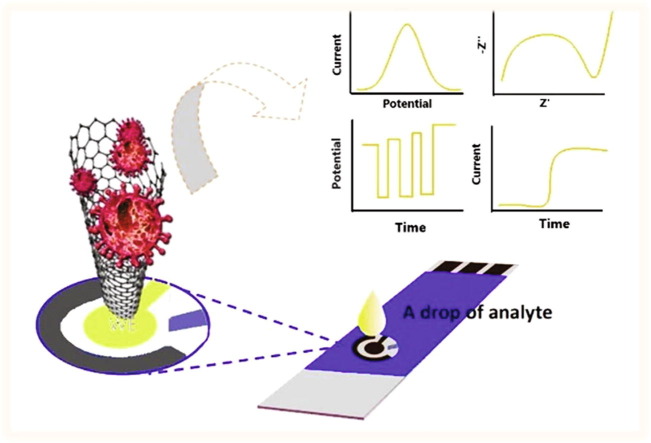
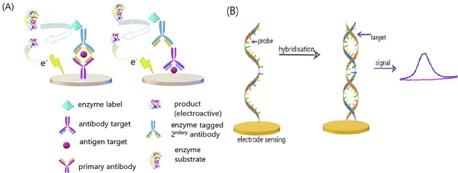
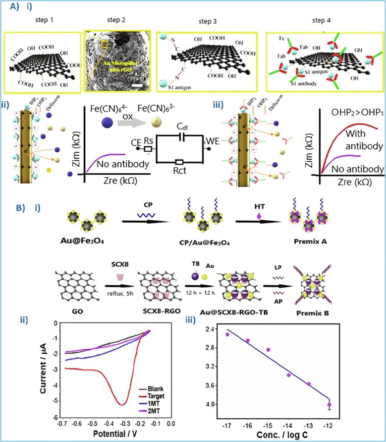
The SARS-CoV-2 virus is still causing a dramatic loss of human lives worldwide, constituting an unprecedented challenge for the society, public health and economy, to overcome. The up-to-date diagnostic tests, PCR, antibody ELISA and Rapid Antigen, require special equipment, hours of analysis and special staff. For this reason, many research groups have focused recently on the design and development of electrochemical biosensors for the SARS-CoV-2 detection, indicating that they can play a significant role in controlling COVID disease. In this review we thoroughly discuss the transducer electrode nanomaterials investigated in order to improve the sensitivity, specificity and response time of the as-developed SARS-CoV-2 electrochemical biosensors. Particularly, we mainly focus on the results appeard on Au-based and carbon or graphene-based electrodes, which are the main material groups recently investigated worldwidely. Additionally, the adopted electrochemical detection techniques are also discussed, highlighting their pros and cos. The nanomaterial-based electrochemical biosensors could enable a fast, accurate and without special cost, virus detection. However, further research is required in terms of new nanomaterials and synthesis strategies in order the SARS-CoV-2 electrochemical biosensors to be commercialized.

Keywords: Au-based nanomaterials; COVID-19 control; Carbon-based nanomaterials; Electrochemical biosensors; Electrochemical detection; SARS-CoV-2 virus detection.


Conflict of interest statement

The authors declare that they have no known competing financial interests or personal relationships that could have appeared to influence the work reported in this paper.

Figures

None
Graphical abstract
Fig. 1
Fig. 1
(Α) Operational principle of a biosensor; (B) Label-free and label-based detection process of biosensors; (C) Label-free aptamer detection process; (D) Antibody-aptamer sandwich type (a&b), aptamers pair sandwich type (c&d). Reproduced with permission.
Fig. 2
Fig. 2
(A) SELEX procedure for aptamers production; (B) Direct (i) and indirect (ii) detection (enzyme-linked immunosorbent assay ELISA) . Reproduced with permission.
Fig. 3
Fig. 3
(A) Antibody-antigen electrochemical immunosensors. (B) DNA-based electrochemical immunosensors. Reproduced with permission.
Fig. 4
Fig. 4
(A) Comparison of LSV measurements in 1 μM DNA solution for albumin (a) and 1-hexanethiol (b) blocking agents on electrode surface . (B) The DNA of SARS genome (oligonucleotides sequence) bound on the modified electrode, producing current . (Ci) Ag reduction current recorded by CV. (Cii) Ag ions, contained in the solution, are reduced into metallic silver (Ag) . CVs of methylene blue covalently attached to a single-stranded DNA (Di) and methylene blue molecule (Dii) on bare (dotted line) and nanostructured screen-printed electrodes (solid line) . (Ε) Comparison of CV response between the dsDNA (blue), dsDNA/rAzu (black), and PSD/rAzu (red). In presence of Ag + ions and oligonucleotides molecules (C = 0.5 pM (10−12 M), rAzu: 0.1 × 10−3g/mL) . Reproduced with permission.
Fig. 5
Fig. 5
(Ai) A schematic illustration of a label-free platform, able to immobilize COVID-19 antibodies on the Au electrodes, coated with RBD SARS-CoV-2 spike-protein; (Aii) Change of the electrical double layer resistance between the Au surface and the RBD bound on it, when antibodies are detected (samples A,B are infected with the virus, while sample C is not) ; (Bi) Au-based SARS-CoV-2 sensor in one chip with three operating modes; (Bii) Comparison of electroanalytical signals between SARS-CoV-2 (2003) cDNA and SARS-CoV2-cDNA ; (C) Electrodeposited Au nanoparticles, where thiol is immobilized/self-assembled and operated as probe for bound the RNA/c-RNA of SARS-CoV-2 ; (Di, Dii) Change in electric properties of a cell-membrane, when the S1 protein (antigen) of SARS-CoV-2 is bound with the immobilized antibody on the Au electrode surface (screen-printed electrode) . Reproduced with permission.
Fig. 6
Fig. 6
(Ai) Functionalized r-GO sheets with carboxylated groups, using a drop-casting process, enriching the r-GO surface with –COOH and –OH groups. (Aii) Schematic representation of the electrical double layer (consisted of Helmholtz planes: OHP & IHP), created between the electrode and the electrolyte. (Aiii) After the targeted antibodies bound with electrode’s antigens, the charge capacitance increases with the increase of the double layer width . (Bi) Schematic illustration of the Au nanoparticles treatment. (Bii) Differential pulse voltammetry (DPV) measurements of the as prepared electrode: i) in blank solution (black), ii) in 10−12 M of the target (red), iii) in 10−12 M of 2 different mismatch artificial targets (MT) (blue and purple). (Biii) Linear relationship (current vs logCanalyte), ranging from 10−17 to 10−12 M . Reproduced with permission.
Fig. 7
Fig. 7
Voltammogram of: (Ai) S-protein detection and (Aii) P-protein detection ; (Bi) TiO2 nanotubes functionalized by Co (Co-TNTs), as electrode material for a SARS-CoV-2 biosensor; (Bii) Linear response for concentration range between 14 nM and 1400 nM and a detection response time of ~ 30 sec; (Ci) Schematic illustration of –NH2 groups of the SARS-CoV-2 proteins or antibodies attached to 1-Pyrenebutyric acid (PBA); (Cii) Current response of real samples detection of the N-proteins, S1-IgG, S1-IgM and CRP ; (Di) FET SARS-CoV-2 biosensor; (Dii) For laboratory sample the lower limit of detection of SARS protein, was recorded at 1 fg/mL and 100 fg/mL . Reproduced with permission.

Similar articles

Cited by

References

    1. Zhou P., Yang X.-L., Wang X.-G., Hu B., Zhang L., Zhang W., Si H.-R., Zhu Y., Li B., Huang C.-L., Chen H.-D., Chen J., Luo Y., Guo H., Jiang R.-D., Liu M.-Q., Chen Y., Shen X.-R., Wang X., Zheng X.-S., Zhao K., Chen Q.-J., Deng F., Liu L.-L., Yan B., Zhan F.-X., Wang Y.-Y., Xiao G.-F., Shi Z.-L. Nature. 2020;579:270–273. - PMC - PubMed
    1. Yuan X., Yang C., He Q., Chen J., Yu D., Li J., Zhai S., Qin Z., Du K., Chu Z., Qin P. ACS Infect. Dis. 2020;6:1998–2016. - PubMed
    1. FDA: U.S Food & Drug, A Closer Look at Coronavirus Disease 2019 (COVID-19) Diagnostic Testing. 2020
    1. Ghaffari A., Meurant R., Ardakani A. Diagnostics. 2020;10(7):453. - PMC - PubMed
    1. Udugama B., Kadhiresan P., Kozlowski H.N., Malekjahani A., Osborne M., Li V.Y.C., Chen H., Mubareka S., Gubbay J.B., Chan W.C.W. ACS Nano. 2020;14:3822–3835. - PubMed

LinkOut - more resources