. 2021 Jun;21(6):458.
doi: 10.3892/ol.2021.12719. Epub 2021 Apr 8.

Inhibition of aquaporins as a potential adjunct to breast cancer cryotherapy

Affiliations

Inhibition of aquaporins as a potential adjunct to breast cancer cryotherapy

Haifa Alkhalifa et al. Oncol Lett. 2021 Jun.

Abstract

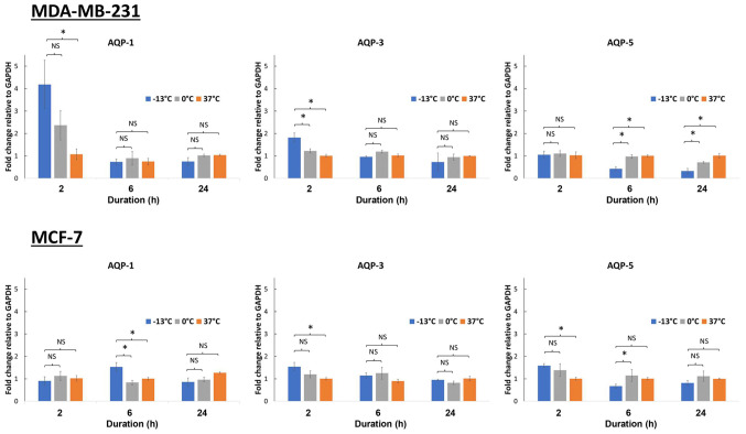
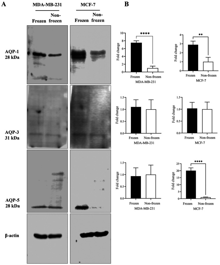
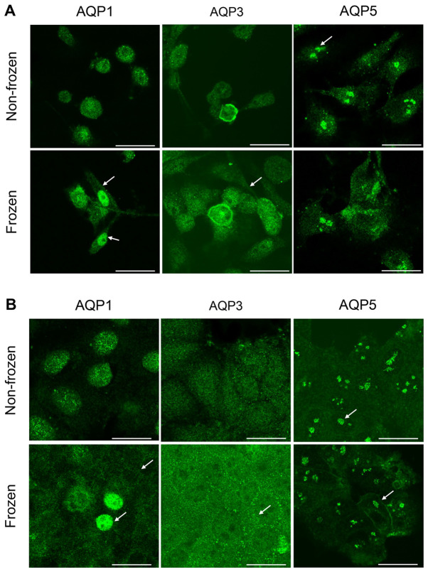
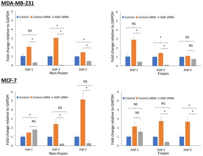
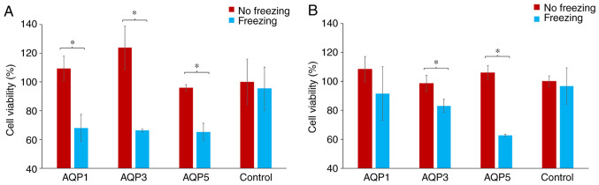
Cryoablation is an emerging type of treatment for cancer. The sensitization of tumors using cryosensitizing agents prior to treatment enhances ablation efficiency and may improve clinical outcomes. Water efflux, which is regulated by aquaporin channels, contributes to cancer cell damage achieved through cryoablation. An increase in aquaporin (AQP) 3 is cryoprotective, whereas its inhibition augments cryodamage. The present study aimed to investigate aquaporin (AQP1, AQP3 and AQP5) gene expression and cellular localization in response to cryoinjury. Cultured breast cancer cells (MDA-MB-231 and MCF-7) were exposed to freezing to induce cryoinjury. RNA and protein extracts were then analyzed using reverse transcription-quantitative PCR and western blotting, respectively. Localization of aquaporins was studied using immunocytochemistry. Additionally, cells were transfected with small interfering RNA to silence aquaporin gene expression and cell viability was assessed using the Sulforhodamine B assay. Cryoinjury did not influence gene expression of AQPs, except for a 4-fold increase of AQP1 expression in MDA-MD-231 cells. There were no clear differences in AQP protein expression for either cell lines upon exposure to frozen and non-frozen temperatures, with the exception of fainter AQP5 bands for non-frozen MCF-7 cells. The exposure of cancer cells to freezing temperatures altered the localization of AQP1 and AQP3 proteins in both MCF-7 and MDA-MD-231 cells. The silencing of AQP1, AQP3 and AQP5 exacerbated MDA-MD-231 cell damage associated with freezing compared with control siRNA. This was also observed with AQP3 and AQP5 silencing in MCF-7 cells. Inhibition of aquaporins may potentially enhance cryoinjury. This cryosensitizing process may be used as an adjunct to breast cancer cryotherapy, especially in the border area targeted by cryoablation where freezing temperatures are not cold enough to induce cellular damage.

Keywords: aquaporins; breast cancer; cryoinjury; cryosensitization; cryotherapy.


Conflict of interest statement

The authors declare that they have no competing interests.

Figures

Figure 1.
Figure 1.
Aquaporin gene expression in response to temperature exposure at −13, 0 and 37°C in 2 breast cancer cell lines (MDA-MB-231 and MCF-7). Following temperature exposure cells were incubated at 37°C for 2, 6 and 24 h. Expression levels of AQP1, AQP3 and AQP5 were then analyzed after 2-, 6- and 24-h periods. *P<0.05. AQP, aquaporin.
Figure 2.
Figure 2.
Effect of the exposure of breast cancer cells to frozen (−13°C) and non-frozen (37°C) temperatures on the expression of AQPs 1, 3 and 5. (A) Total cell lysates were analyzed by western blotting using AQP-1, 3, and 5 antibodies and β-actin as a loading control. (B) Quantification of the AQP-1, 3, and 5 normalized to β-actin and expressed as fold of change. **P<0.05 and ****P<0.001. AQP, aquaporin.
Figure 3.
Figure 3.
Representative results of immunocytochemistry of aquaporin proteins (AQP1, AQP3 and AQP5) in (A) MDA-MB-231 and (B) MCF-7 cells. Upper panels show cells not exposed to freezing temperatures while lower panels show cells that were frozen at −13°C. (A) Intense staining of AQP1 and AQP3 was seen within the plasma and nuclear membranes following freezing for MDA-MB-231 cells (Fig. 3A, arrows); indicating relocation of AQP1 and AQP3 to membranes. Similar staining for AQP3 expression was observed in the plasma membrane following the freezing of MDA-MB-231 cells (Fig. 3A, arrows). AQP5 protein expression was clustered and localized in the nucleus in both treatment conditions (Fig. 3A, arrows). (B) In MCF-7 cells AQP1 and AQP3 showed localization in membranes following freezing. AQP1 had a diffused cytoplasm staining and accumulated in the plasma membrane compared with AQP1 localization in non-frozen cells (Fig. 3B, arrows); few cells had intense nuclear staining (Fig. 3B, arrows). Plasma membrane localization of AQP5 in MCF-7 cells appeared more intense following exposure to freezing temperature (Fig. 3B, arrows). There were no discernible changes in staining and localization of AQP5 in both MDA-MB-231 and MCF-7 cells in response to freezing. Scale bar, 25 µm. AQP, aquaporin.
Figure 4.
Figure 4.
Assessment of gene knockdown following small siRNA transfection. AQP gene silencing was assessed using RT-qPCR in MDA-MB-231 and MCF-7 breast cancer cells exposed to frozen and non-frozen conditions. siRNA targeting AQP1, AQP3 and AQP5 were used to knock down gene expression. In addition, untreated cells were used as a control as well as control-siRNA. Following transfection of cells with siRNA, cells were cryoinjured for 10 min at −13°C. Gene expression was then assessed by RT-qPCR relative to the housekeeping gene GAPDH. *P<0.05, Si, small interfering; RT-q, reverse transcription-quantitative; AQP, aquaporin.
Figure 5.
Figure 5.
Assessment of cell viability following siRNA transfection and exposure to freezing in MDA-MB-231 and MCF-7 breast cancer cells. (A) MDA-MB-231 and (B) MCF-7 cells. Cell viability was assayed using the Sulforhodamine B assay following transfection with siRNA and exposure to freezing and non-freezing conditions. Cells were transfected with siRNA targeting 3 aquaporins: AQP1, AQP3 and AQP5. The control group was the scrambled negative-control siRNA. *P<0.05. Si, small interfering; AQP, aquaporin.

Similar articles

Cited by

References

    1. Bray F, Ferlay J, Soerjomataram I, Siegel RL, Torre LA, Jemal A. Global cancer statistics 2018: GLOBOCAN estimates of incidence and mortality worldwide for 36 cancers in 185 countries. CA Cancer J Clin. 2018;68:394–424. doi: 10.3322/caac.21492. - DOI - PubMed
    1. Global Burden of Disease Cancer Collaboration. Fitzmaurice C, Allen C, Barber RM, Barregard L, Bhutta ZA, Brenner H, Dicker DJ, Chimed-Orchir O, Dandona R, et al. Global, regional, and national cancer incidence, mortality, years of life lost, years lived with disability, and disability-adjusted life-years for 32 cancer groups, 1990 to 2015: A systematic analysis for the global burden of disease study. JAMA Oncol. 2017;3:524–548. doi: 10.1001/jamaoncol.2016.5688. - DOI - PMC - PubMed
    1. PDQ Adult Treatment Editorial Board, corp-author. PDQ Cancer Information Summaries. National Cancer Institute (USA); Bethesda, MD: 2002. Breast cancer treatment (Adult) (PDQ®): Patient version.
    1. Sharma GN, Dave R, Sanadya J, Sharma P, Sharma KK. Various types and management of breast cancer: An overview. J Adv Pharm Technol Res. 2010;1:109–126. - PMC - PubMed
    1. Pusceddu C, Melis L, Ballicu N, Meloni P, Sanna V, Porcu A, Fancellu A. Cryoablation of primary breast cancer in patients with metastatic disease: Considerations arising from a single-centre data analysis. Biomed Res Int. 2017;2017:3839012. doi: 10.1155/2017/3839012. - DOI - PMC - PubMed