. 2021 Jun;21(6):472.
doi: 10.3892/ol.2021.12733. Epub 2021 Apr 13.

Long non-coding RNA SNHG17 enhances the aggressiveness of C4-2 human prostate cancer cells in association with β-catenin signaling

Affiliations

Long non-coding RNA SNHG17 enhances the aggressiveness of C4-2 human prostate cancer cells in association with β-catenin signaling

Haijun Zhao et al. Oncol Lett. 2021 Jun.

Abstract

Long non-coding (lnc) RNAs have emerged as important regulators of cancer development and progression. Several lncRNAs have been reported to be associated with prostate cancer (PCa); however, the involvement of lncRNA SNHG17 in PCa remains unclear. In the present study, the mRNA expression level of SNHG17 in 58 pairs of PCa tumor samples and adjacent non-tumor tissues, as well as in PCa tumor cell lines was analyzed. The regulatory effect of SNHG17 on the oncogenic phenotypes of the C4-2 tumor cell line was also investigated. The clinicopathological analysis revealed that SNHG17 mRNA expression level was increased in the PCa tumor samples, and its high expression levels were associated with poor patient outcomes, indicating that SNHG17 may act as a biomarker for the prognosis of PCa. SNHG17 mRNA expression level was also increased in different PCa tumor cell lines. Functionally, SNHG17 increased C4-2 tumor cell growth and aggressiveness by stimulating tumor cell proliferation, survival, invasion and resistance to chemotherapy. Furthermore, SNHG17 promoted in vivo tumor growth in a xenograft mouse model. Notably, the SNHG17-induced in vitro and in vivo oncogenic effects were associated with activation of the β-catenin pathway. The results from the present study revealed that lncRNA SNHG17 could be an important regulator in the oncogenic properties of human PCa and may; therefore, represent a potential PCa therapeutic target.

Keywords: SNHG17; Wnt; lncRNA; prostate cancer.


Conflict of interest statement

The authors declare that they have no competing interests.

Figures

Figure 1.
Figure 1.
Upregulation of lncRNA SNHG17 is associated with poor outcomes in patients with PCa. (A) Reverse transcription-quantitative PCR was performed to determine the SNHG17 mRNA expression level in 58 paired tumor and non-tumor tissues, and (B) HPrEC, LNCaP and C4-2 cell lines (n=3). (C) Kaplan-Meier survival curve indicating that patients with high SNHG17 mRNA expression levels had poorer overall survival rates compared with patients with low expression levels. The data are presented as the mean ± SD. *P<0.05, **P<0.01. PCa, prostate cancer; lnc, long non-coding.
Figure 2.
Figure 2.
KD of SNHG17 inhibits the proliferation and survival but increases caspase 3 activity of the C4-2 tumor cells. (A) Reverse transcription-quantitative PCR was used to determine the mRNA expression level of SNHG17 in untransduced, SNHG17-KD and control cells (n=3). (B) MTT assay was used to determine cell proliferation in cells transfected with SNHG17 or control shRNA (n=4). (C) Viability assay was used to measure cell survival cells transduced with SNHG17 or control shRNA (n=3). (D) Caspase 3 activity was measured in cells transduced with SNHG17 or control shRNA (n=3). The data are presented as the mean ± SD. *P<0.05, **P<0.01. FCS, fetal calf serum; KD, knockdown; OD, optical density; sh, short hairpin; RLU, relative luminescence units.
Figure 3.
Figure 3.
KD of SNHG17 suppresses the invasion and chemotherapeutic resistance of the C4-2 tumor cells. (A) Invasion assay showing that KD of SNHG17 reduces the number of invasive cells (n=3); magnification ×400. (B) Viability assay in control and SNHG17-KD tumor cells, treated with docetaxel (0, 1, 2.5, 5 and 10 nM) for 1, 3 and 5 days, respectively. (n=4). The data are presented as the mean ± SD. *P<0.05, **P<0.01. KD, knockdown.
Figure 4.
Figure 4.
KD of SNHG17 suppresses in vivo tumor growth. A total of 14 BALB/c nude mice were divided into two groups (7 mice/group) and were injected subcutaneously with 1×10⁶ control or SNHG17-KD C4-2 tumor cells. (A) Mean tumor size. (B) Images of the tumor samples harvested at day 50 following implantation. Scale bar, 1 cm. (C) Mean tumor weight. (D-a) Immunohistochemistry Ki-67 staining in tumors. Scale bar, 100 µm (D-b) Quantification of the Ki-67 positive cells. The data are presented as the mean ± SD. *P<0.05, **P<0.01. KD, knockdown.
Figure 5.
Figure 5.
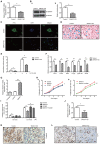
SNHG17 facilitates C4-2 tumor growth via β-catenin. (A) RT-qPCR and (B-a) western blot analysis revealed that β-catenin mRNA and protein expression level was decreased in the SNHG17-KD cells, respectively (n=3). (B-b) Densitometry analysis of β-catenin (normalized to β-tubulin using ImageJ software). (C) Immunofluorescence staining of β-catenin and (D) immunocytochemical staining of TCF1 in the control or SNHG17-KD tumor cells. (E) TCF reporter assay in the control or SNHG17-KD cells, treated with/without LiCl (5 mM) for 24 h (n=3). RT-qPCR was performed to determine the mRNA expression level of (F) different genes in the control or SNHG17-KD tumor cells and (G) SNHG17 in cells transfected with pcDNA-SNHG17 vector (n=3). MTT assay was used to determine cell proliferation in the (H-a) control, SNHG17-overexpressing and control cells treated with ICG001 (10 µM) and (H-b) in SNHG17-overexpressing cells treated with/without ICG001 (n=4). (I) Caspase 3 activity assay was measured in the control or SNHG17-KD cells, treated with/without ICG001 (10 µM) for 48 h (n=3). (J) RT-qPCR was used to measure the mRNA expression level of β-catenin in the tumor samples (n=3). Immunohistochemistry staining of (K) β-catenin and (L) TCF1 in the tumor samples. For C: Scale bar, 50 µm; For D, K and L: Scale bar, 100 µm. The data are presented as the mean ± SD. *P<0.05, **P<0.01. RT-qPCR, reverse transcription-quantitative PCR; KD, knockdown; OD, optical density.

Similar articles

Cited by

References

    1. Global Burden of Disease Cancer Collaboration. Fitzmaurice C, Allen C, Barber RM, Barregard L, Bhutta ZA, Brenner H, Dicker DJ, Chimed-Orchir O, Dandona R, et al. Global, regional, and national cancer incidence, mortality, years of life lost, years lived with disability, and disability-adjusted life-years for 32 cancer groups, 1990 to 2015: A systematic analysis for the global burden of disease study. JAMA Oncol. 2017;3:524–548. doi: 10.1001/jamaoncol.2016.5688. - DOI - PMC - PubMed
    1. Chandrasekar T, Yang JC, Gao AC, Evans CP. Mechanisms of resistance in castration-resistant prostate cancer (CRPC) Transl Andro Urol. 2015;4:365–380. - PMC - PubMed
    1. Mansinho A, Macedo D, Fernandes I, Costa L. Castration-Resistant prostate cancer: Mechanisms, targets and treatment. Adv Exp Med Biol. 2018;1096:117–133. doi: 10.1007/978-3-319-99286-0_7. - DOI - PubMed
    1. Aird J, Baird AM, Lim MCJ, McDermott R, Finn SP, Gray SG. Carcinogenesis in prostate cancer: The role of long non-coding RNAs. Noncoding RNA Res. 2018;3:29–38. doi: 10.1016/j.ncrna.2018.01.001. - DOI - PMC - PubMed
    1. Weiss M, Plass C, Gerhauser C. Role of lncRNAs in prostate cancer development and progression. Biol Chem. 2014;395:1275–1290. doi: 10.1515/hsz-2014-0201. - DOI - PubMed