Pilonidal Cyst Excision: Primary Midline Closure with versus without Closed Incision Negative Pressure Therapy
- PMID: 33907657
- PMCID: PMC8062152
- DOI: 10.1097/GOX.0000000000003473
Pilonidal Cyst Excision: Primary Midline Closure with versus without Closed Incision Negative Pressure Therapy
Abstract
Background: Pilonidal cysts are a painful condition that primarily affect young adult men. In the literature, numerous operative techniques for resolving pilonidal cysts are described, with variable outcomes. The objective of this study was to compare primarily closed midline incisions managed with or without the use of closed incision negative pressure therapy after pilonidal cyst excision.
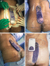
Methods: Twenty-one patients underwent excision and midline primary closure. Postoperative care composed of closed incisional negative pressure therapy (study group; n = 10) or gauze dressings (control group; n = 11). In both groups, the sutures were partially removed on day 14 and completely removed on day 21. Compared outcomes included the duration of hospitalization, pain on the day of surgical procedure, and on postoperative day 7, and time-to-healing.
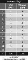
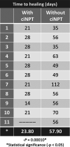
Results: The median hospital stay was about 9 hours and 23 hours in the study and control groups, respectively (P < 0.05). The median pain scores on the day of operation were 1.20/10 in the study group and 3.36/10 in the control group (P < 0.05). On day 7, study group showed median pain score 0.9/10 and control group showed 2.63/10 (P < 0.05). The mean healing time was 23.8 and 57.9 days in the ciNPT group and gauze group, respectively (P < 0.05).
Conclusion: These outcomes supported the incorporation of closed incision negative pressure therapy into our surgical treatment protocol for pilonidal cysts.
Copyright © 2021 The Authors. Published by Wolters Kluwer Health, Inc. on behalf of The American Society of Plastic Surgeons.
Conflict of interest statement
Disclosure: The authors have no financial interest to declare in relation to the content of this article.
Figures


References
-
- da Silva JH. Pilonidal cyst: Cause and treatment. Dis Colon Rectum. 2000; 43:1146–1156. - PubMed
-
- Elalfy K, Emile S, Lotfy A, et al. Bilateral gluteal advancement flap for treatment of recurrent sacrococcygeal pilonidal disease: A prospective cohort study. Int J Surg. 2016; 29:1–8. - PubMed
-
- Aysan E, Ilhan M, Bektas H, et al. Prevalence of sacrococcygeal pilonidal sinus as a silent disease. Surg Today. 2013; 43:1286–1289. - PubMed
-
- Karydakis GE. Easy and successful treatment of pilonidal sinus after explanation of its causative process. Aust N Z J Surg. 1992; 62:385–389. - PubMed
LinkOut - more resources
Full Text Sources
Other Literature Sources
