. 2021 Jul;24(1):485.
doi: 10.3892/mmr.2021.12124. Epub 2021 Apr 28.

Non-toxic sulfur inhibits LPS-induced inflammation by regulating TLR-4 and JAK2/STAT3 through IL-6 signaling

Affiliations

Non-toxic sulfur inhibits LPS-induced inflammation by regulating TLR-4 and JAK2/STAT3 through IL-6 signaling

Dong Young Kang et al. Mol Med Rep. 2021 Jul.

Abstract

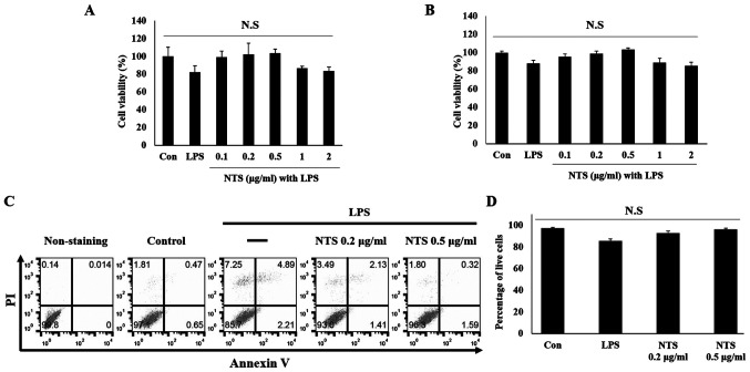
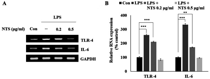
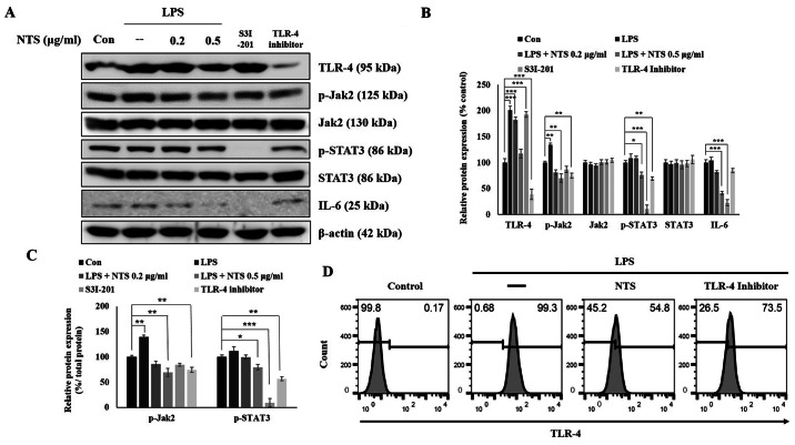
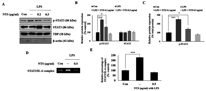
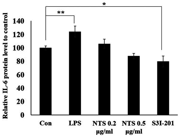
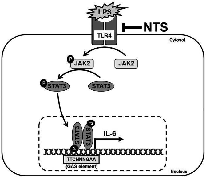
Janus kinase 2 (JAK2) and STAT3 signaling is considered a major pathway in lipopolysaccharide (LPS)‑induced inflammation. Toll‑like receptor 4 (TLR‑4) is an inflammatory response receptor that activates JAK2 during inflammation. STAT3 is a transcription factor for the pro‑inflammatory cytokine IL‑6 in inflammation. Sulfur is an essential element in the amino acids and is required for growth and development. Non‑toxic sulfur (NTS) can be used in livestock feeds as it lacks toxicity. The present study aimed to inhibit LPS‑induced inflammation in C2C12 myoblasts using NTS by regulating TLR‑4 and JAK2/STAT3 signaling via the modulation of IL‑6. The 3‑(4,5‑dimethylthiazol‑2‑yl)‑2,5‑diphenyltetrazolium bromide assay was conducted to analyze cell viability and reverse transcription polymerase chain reaction and western blotting performed to measure mRNA and protein expression levels. Chromatin immunoprecipitation and enzyme‑linked immunosorbent assays were used to determine the binding activity of proteins. The results indicated that NTS demonstrated a protective effect against LPS‑induced cell death and inhibited LPS‑induced expression of TLR‑4, JAK2, STAT3 and IL‑6. In addition, NTS inhibited the expression of nuclear phosphorylated‑STAT3 and its binding to the IL‑6 promoter. Therefore, NTS may be a potential candidate drug for the treatment of inflammation.

Keywords: IL-6; Janus kinase 2; STAT3; Toll-like receptor 4; non-toxic sulfur.


Conflict of interest statement

Hyoung Do Kim is affiliated with Nara Bio Co., Ltd., which provided funding for this study and supplied non-toxic sulfur. The remaining authors declare that they have no competing interests.

Figures

Figure 1.
Figure 1.
Effect of NTS on the viability of LPS-treated C2C12 cells. (A) MTT assay was performed to evaluate viability of C2C12 cells following treatment with or without 100 ng/ml LPS or 0.1–2 µg/ml NTS for 24 h. Cell viability was determined using the formula % viability = (fluorescence value of methylsulfonylmethane/fluorescence value of non-treated control) ×100. Data are representative of three independent experiments. Non-significant results were observed via statistical analysis. (B) WST-1 assay showing the cyto protective effects of NTS against LPS-induced cell death. Data are representative of three independent experiments. Non-significant results were observed via statistical analysis. (C) Flow cytometry analysis using Annexin V and PI staining in C2C12 cells following treatment with LPS and NTS for 24 h. (D) Graphical representation of the percentage of cells undergoing phases of apoptosis following treatment with 100 ng/ml LPS or 0.2 and 0.5 µg/ml NTS in C2C12 myoblasts. The values are presented as mean ± standard error of the mean of three independent experiments performed in triplicate (n=3). NTS, non-toxic sulfur; LPS, lipopolysaccharide; Con, control; N.S., non-significant.
Figure 2.
Figure 2.
NTS inhibits TLR-4 and IL-6 mRNA expression. (A) Reverse transcription-semi-quantitative PCR results showing the analysis of TLR-4 and IL-6 mRNA expression following the treatment of C2C12 cells with 100 ng/ml LPS or 0.2 and 0.5 µg/ml NTS for 24 h. (B) Relative mRNA expression levels of TLR-4 and IL-6 were determined via densitometry and were normalized to GAPDH mRNA. Data are representative of three independent experiments. **P<0.01 and ***P<0.001 vs. control. NTS, non-toxic sulfur; TLR-4, Toll-like receptor 4; LPS, lipopolysaccharide; Con, control.
Figure 3.
Figure 3.
NTS inhibits TLR-4, JAK2, STAT3 and IL-6 signaling. (A) Western blotting showing the expression of TLR-4, JAK2, p-JAK2, STAT3, p-STAT3 and IL-6 proteins following the treatment of C2C12 cells with 100 ng/ml LPS and 0.2 or 0.5 µg/ml NTS or 100 µM S3I-201 or 80 µM TLR4-C34 for 24 h. (B) Relative protein expression levels of TLR-4, JAK2, p-JAK2, STAT3, p-STAT3 and IL-6 were determined via densitometry and were normalized to β-actin. Data are representative of three independent experiments. (C) Relative expression levels of p-JAK2 and p-STAT3 were determined via densitometry and were normalized to total JAK2 and STAT3. (D) Flow cytometry (fluorescence-activated cell sorting) analysis of TLR-4 expression in C2C12 cells showing the expression levels of LPS-induced TLR-4 and its inhibition by 0.5 µg/ml NTS or 80 µM Toll-like receptor 4 inhibitor (TLR4-C34) for 24 h. *P<0.05, **P<0.01 and ***P<0.001 vs. control. NTS, non-toxic sulfur; TLR-4, Toll-like receptor 4; JAK2, Janus kinase 2; p-, phosphorylated; LPS, lipopolysaccharide; Con, control.
Figure 4.
Figure 4.
NTS inhibits the expression levels of nuclear p-STAT3 and its binding to the IL-6 promoter. (A) Western blotting of the nuclear extracts of C2C12 cells showing the expression of STAT3 and p-STAT3 nuclear proteins following treatment with 100 ng/ml LPS and 0.2 or 0.5 µg/ml NTS for 24 h. (B) Relative expression levels of STAT3 and p-STAT3 nuclear proteins were determined via densitometry and normalized to TBP. Data are representative of three independent experiments. (C) Relative expression levels of nuclear p-STAT3 determined via densitometry and were normalized to total STAT3. (D) ChIP analysis of STAT3-IL-6 complex formation in C2C12 cells following treatment with 100 ng/ml LPS or 0.5 µg/ml NTS for 24 h. Data were quantified using reverse transcription-semi-quantitative PCR. (E) Relative STAT3-IL-6 complex binding obtained in C2C12 cells with 100 ng/ml LPS or 0.5 µg/ml NTS for 24 h by ChIP assay and expressed as a percentage of the control. Data are representative of three independent experiments. *P<0.05 and ***P<0.001 vs. control. NTS, non-toxic sulfur; p-, phosphorylated; LPS, lipopolysaccharide; ChIP, chromatin immunoprecipitation; Con, control; TBP, Tata-binding protein.
Figure 5.
Figure 5.
NTS inhibits STAT3-dependent IL6 expression. ELISA analysis showing the inhibition of LPS-induced expression of IL-6 following treatment with 0.2 and 0.5 µg/ml of NTS or 100 µM S3I-201 for 24 h. Data are representative of three independent experiments. *P<0.05 and **P<0.01 vs. control. NTS, non-toxic sulfur; Con, control; LPS, lipopolysaccharide.
Figure 6.
Figure 6.
Molecular regulatory mechanism of LPS-induced inflammatory responses by TLR-4 and JAK2/STAT3 via IL-6 signaling. The anti-inflammatory activities of NTS are mediated by inhibiting these pathways and the binding of STAT3 to the promoter of the pro-inflammatory cytokine IL-6. LPS, lipopolysaccharide; NTS, non-toxic sulfur; TLR-4, Toll-like receptor 4; JAK2, Janus kinase 2; p-, phosphorylated; GAS, γ-interferon activation site.

Similar articles

Cited by

References

    1. Chen L, Deng H, Cui H, Fang J, Zuo Z, Deng J, Li Y, Wang X, Zhao L. Inflammatory responses and inflammation-associated diseases in organs. Oncotarget. 2017;9:7204–7218. doi: 10.18632/oncotarget.23208. - DOI - PMC - PubMed
    1. Catorce MN, Gevorkian G. LPS-induced murine neuroinflammation model: Main features and suitability for pre-clinical assessment of nutraceuticals. Curr Neuropharmacol. 2016;14:155–164. doi: 10.2174/1570159X14666151204122017. - DOI - PMC - PubMed
    1. Heinbockel L, Weindl G, Martinez-de-Tejada G, Correa W, Sanchez-Gomez S, Bárcena-Varela S, Goldmann T, Garidel P, Gutsmann T, Brandenburg K. Inhibition of lipopolysaccharide- and lipoprotein-induced inflammation by antitoxin peptide Pep19-2.5. Front Immunol. 2018;9:1704. doi: 10.3389/fimmu.2018.01704. - DOI - PMC - PubMed
    1. Kawai T, Akira S. Signaling to NF-kappaB by Toll-like receptors. Trends Mol Med. 2007;13:460–469. doi: 10.1016/j.molmed.2007.09.002. - DOI - PubMed
    1. Liu T, Zhang L, Joo D, Sun SC. NF-κB signaling in inflammation. Signal Transduct Target Ther. 2017;2:2. doi: 10.1038/sigtrans.2017.23. - DOI - PMC - PubMed

MeSH terms