

Tissue distribution of epirubicin after severe extravasation in humans
- PMID: 33907881
- PMCID: PMC8236455
- DOI: 10.1007/s00280-021-04280-8
Tissue distribution of epirubicin after severe extravasation in humans
Abstract
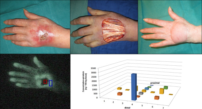
Purpose: As critical parameter after extravasation of cytotoxic vesicants, anthracyclines were determined in removed tissue from patients requiring surgical intervention due to tissue necrosis. We monitored their distribution within the affected lesion to establish a possible dose-toxicity relation.
Methods: From six patients scheduled for surgery, removed tissue flaps were systematically analysed by HPLC (epirubicin: 5 subjects; doxorubicin: 1 subject).
Results: After extravasation, tissue concentrations were highly variable with an individual anthracycline distribution pattern ranging from a few nanograms up to 17 µg per 100 mg tissue, which indicated a substantial difference in tissue sensitivity among patients. The resection borders coincided with the extension of the erythema and guided the surgical intervention after demarcation of the lesion, which occurred usually 2 or 3 weeks after extravasation. At that time, drug was hardly detected at the resection borders. Wound drains were negative for the extravasated drugs while showing a time profile of vascular growth factors and inflammatory cytokines, which was highly similar to routine surgery. In all six patients, surgical debridement with immediate wound closure led to healing within approximately 2 weeks, when therapy was resumed in all patients with reasonable time delay.
Conclusion: Surgical intervention after demarcation of the extravasation lesion allows for almost uninterrupted continuation of treatment independent of the amount of extravasated anthracycline. As even minor amounts of the vesicants may trigger tissue necrosis, preventive measures merit the highest priority.
Keywords: Anthracycline; Extravasation; HPLC; Surgery; Tissue concentration.
Conflict of interest statement
The authors declare no conflicts of interest.
Figures

References
-
- Mader I, Furst-Weger P, Mader RM, Nogler-Semenitz E, Wassertheurer S. Extravasation of cytotoxic agents. 2. New York: Springer; 2010. - PubMed
Publication types
MeSH terms
Substances
LinkOut - more resources
Full Text Sources
Other Literature Sources
Research Materials
