. 2021 Dec;529(17):3751-3771.
doi: 10.1002/cne.25163. Epub 2021 May 6.

The synaptic inputs and thalamic projections of two classes of layer 6 corticothalamic neurons in primary somatosensory cortex of the mouse

Affiliations

The synaptic inputs and thalamic projections of two classes of layer 6 corticothalamic neurons in primary somatosensory cortex of the mouse

Courtney Michelle Whilden et al. J Comp Neurol. 2021 Dec.

Erratum in

  • Erratum.
    [No authors listed] [No authors listed] J Comp Neurol. 2022 May;530(7):1126. doi: 10.1002/cne.25315. J Comp Neurol. 2022. PMID: 35338485 Free PMC article. No abstract available.

Abstract

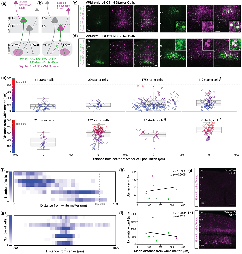
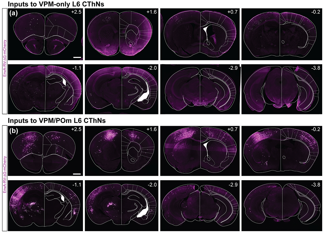
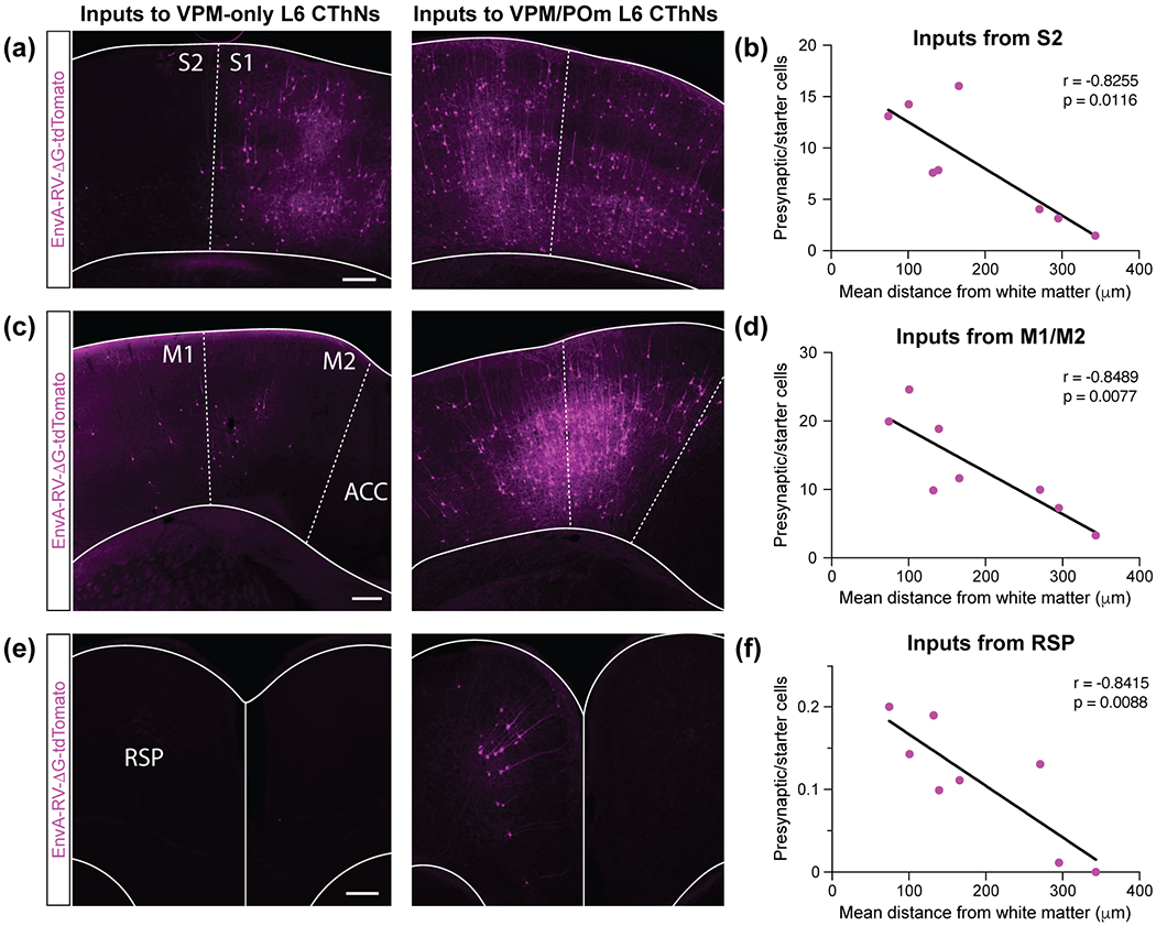
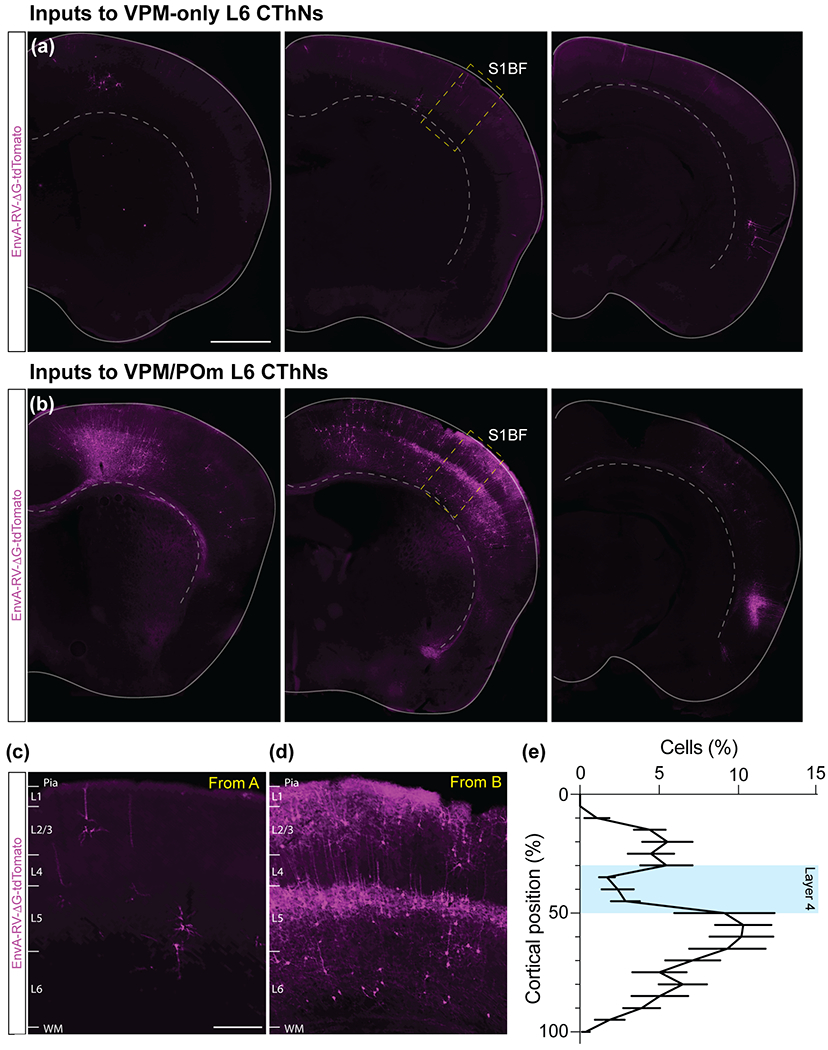
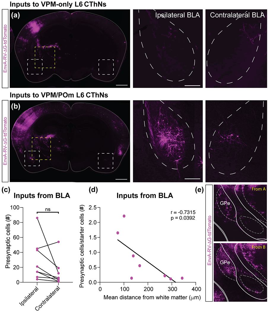
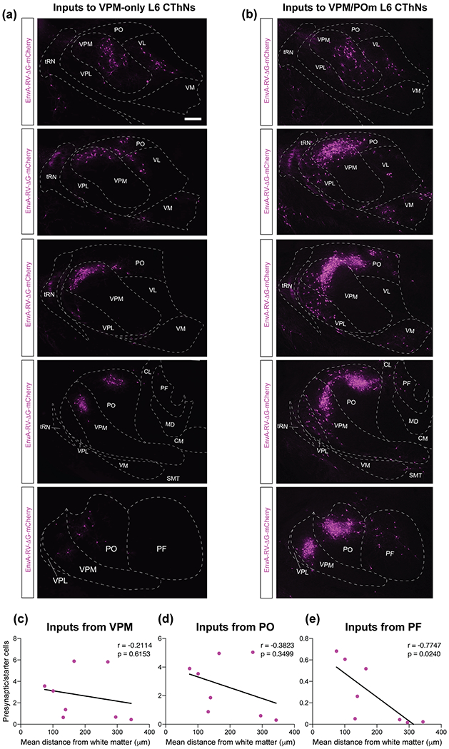
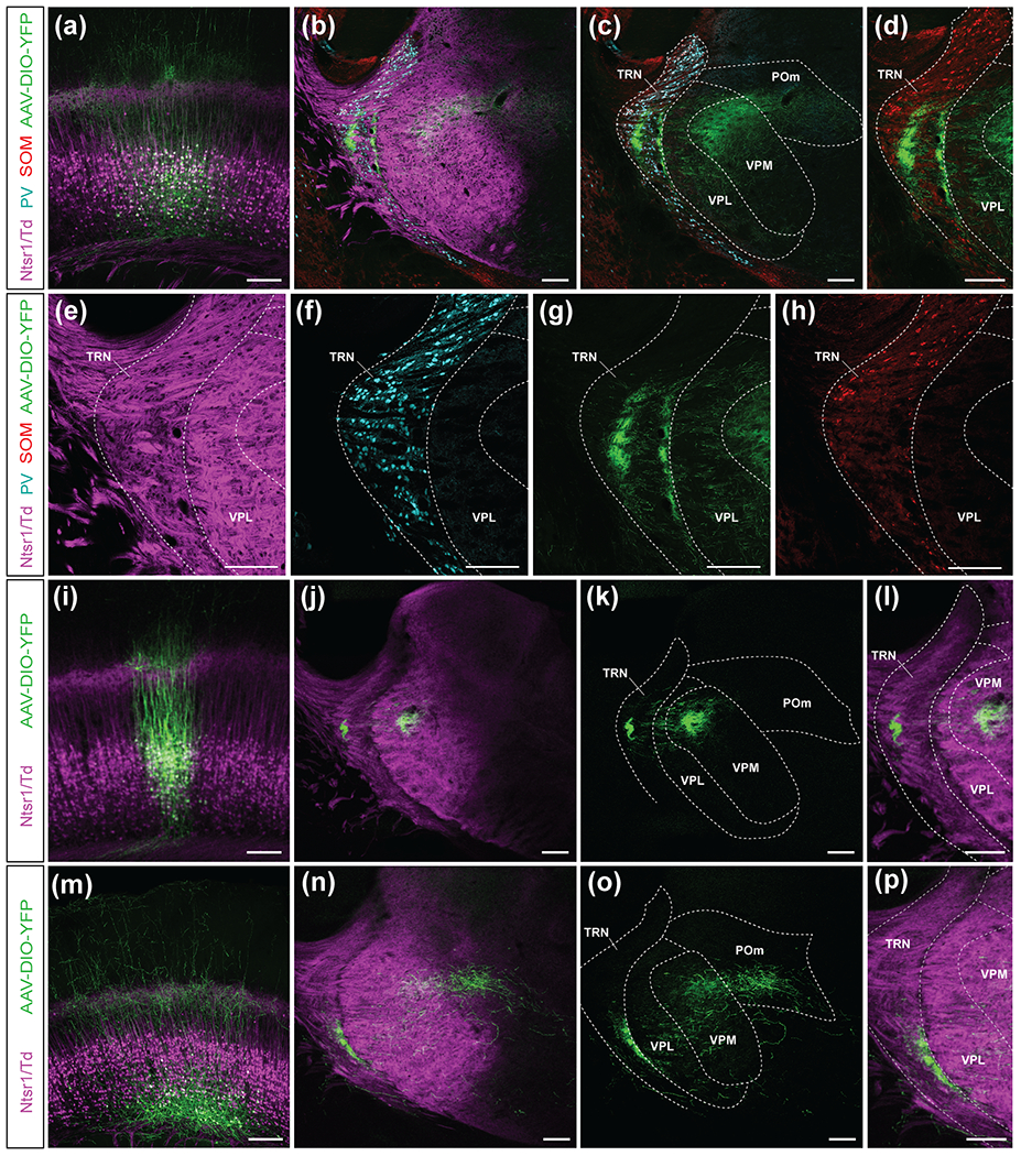
Although corticothalamic neurons (CThNs) represent the largest source of synaptic input to thalamic neurons, their role in regulating thalamocortical interactions remains incompletely understood. CThNs in sensory cortex have historically been divided into two types, those with cell bodies in Layer 6 (L6) that project back to primary sensory thalamic nuclei and those with cell bodies in Layer 5 (L5) that project to higher-order thalamic nuclei and subcortical structures. Recently, diversity among L6 CThNs has increasingly been appreciated. In the rodent somatosensory cortex, two major classes of L6 CThNs have been identified: one projecting to the ventral posterior medial nucleus (VPM-only L6 CThNs) and one projecting to both VPM and the posterior medial nucleus (VPM/POm L6 CThNs). Using rabies-based tracing methods in mice, we asked whether these L6 CThN populations integrate similar synaptic inputs. We found that both types of L6 CThNs received local input from somatosensory cortex and thalamic input from VPM and POm. However, VPM/POm L6 CThNs received significantly more input from a number of additional cortical areas, higher order thalamic nuclei, and subcortical structures. We also found that the two types of L6 CThNs target different functional regions within the thalamic reticular nucleus (TRN). Together, our results indicate that these two types of L6 CThNs represent distinct information streams in the somatosensory cortex and suggest that VPM-only L6 CThNs regulate, via their more restricted circuits, sensory responses related to a cortical column while VPM/POm L6 CThNs, which are integrated into more widespread POm-related circuits, relay contextual information.

Keywords: corticothalamic neurons; layer 6; rabies virus; somatosensory cortex; thalamic reticular nucleus; trans-synaptic tracing.


Conflict of interest statement

Competing financial interests: The authors declare no competing financial interests.

Figures

Figure 1
Figure 1
Summary of experimental approach for identifying presynaptic inputs to different classes of layer 6 corticothalamic neurons (L6 CThNs). (a,b) On Day 1, stereotactic injections of two helper AAV viruses encoding rabies G protein and the receptor TVA respectively were biased to upper L6a (a) or lower L6a (b) to enrich transfections for VPM-only (a) or VPM/POm (b) L6 CThNs. Two weeks later, an EnvA-pseudotyped G-deleted rabies virus was injected into VPM (a) or POm (b) to infect VPM-projecting or POm-projecting L6 CThNs. (c,d) Lower (left three panels) and higher (right three panels) magnification images of the starter cell population for an experiment enriched for VPM-only L6 CThNs (c) or VPM/POm L6 CThNs (d). The dashed white boxes indicate the regions shown at higher magnification in the right three panels. Starter cells co-express tdTomato (magenta) due to the rabies virus and either Blue Fluorescent Protein (BFP, green) or enhanced Green Fluorescent Protein (eGFP, green) due to the AAV TVA helper virus as shown in the higher magnification insets. (e) The location of each double-labeled starter cell for the eight experiments is shown. The box plots represent quartiles centered on the median laminar position of each starter cell population. Outliers more than 1.5 time the interquartile range are also shown. Higher magnification images from the experiment marked with (#, 86 starter cells) are shown in Figure 2a and 3c, left; the experiment marked with ($, 112 starter cells) in Figure 2b, 3a, right, 3c, right, 3e, right, 5b; and the experiment marked with (@; 23 starter cells) in Figure 3a, left, 3e, left and 5a. Summary of the vertical (f) and horizontal (g) distribution of the starter cell population for the eight experiments. (h) The total number of starter cells in each experiment did not correlate with the mean laminar position of the starter cell population (r = 0.1682, p = 0.6905, n = 8 mice). (i) The horizontal extent of each starter cell population did not correlate with the mean laminar position of the starter cell population (r = −0.2372, p = 0.5716, n = 8 mice). (j) Example control experiment showing the lack of rabies infection in the absence of TVA expression. (k) Example control experiment showing the lack of trans-synaptic spread of the rabies in the absence of G protein expression. Scale bars: (c,d,j,k) = 200 μm, insets in (c,d) = 10 μm.
Figure 2
Figure 2
Distribution of tdTomato-expressing neurons in the brains from two example experiments. (a,b) Eight coronal sections spanning the anterior-posterior axis for an experiment with a starter cell population enriched for VPM-only L6 CThNs (a) or VPM/POm L6 CThNs (b). The experiment in (a) corresponds to the experiment marked with a (#) in Figure 1e with 86 starter cells and the experiment in (b) corresponds to the experiment marked with a ($) with 112 starter cells. The starter cells were located in the hemisphere on the left in all images. Schematics of major landmarks were overlaid on the images. Scale bars: (a,b) = 1 mm.
Figure 3
Figure 3
VPM/POm layer 6 corticothalamic neurons (L6 CThNs) receive more input from ipsilateral secondary somatosensory cortex (S2), cortical motor areas and retrosplenial cortex than VPM-only L6 CThNs. (a) Images of coronal sections of primary sensory cortex (S1) and S2 for an example experiment with a starter cell population enriched for VPM-only (left) or VPM/POm L6 CThNs (right). (b) Correlation between the mean laminar position of the starter cell population and the normalized number of tdTomato-positive cells in ipsilateral S2 (r = −0.8255, p = 0.0116, n = 8 mice). (c) Images of coronal sections of primary and secondary motor cortex (M1/M2) for an example experiment with a starter cell population enriched for VPM-only (left) or VPM/POm L6 CThNs (right). (d) Correlation between the mean laminar position of the starter cell population and the normalized number of tdTomato-positive cells in ipsilateral M1/M2 (r = −0.8489, p = 0.0077, n = 8 mice). (e) Images of coronal sections of retrosplenial cortex (RSP) for an example experiment with a starter cell population enriched for VPM-only (left) or VPM/POm L6 CThNs (right). (f) Correlation between the mean laminar position of the starter cell population and the normalized number of tdTomato-positive cells in ipsilateral RSP (r = −0.8415, p = 0.0088, n = 8 mice). Dashed lines indicate areal boundaries. Solid line indicates boundary between the hemispheres. Scale bars: (a,c,e) = 200 μm.
Figure 4
Figure 4
VPM/POm layer 6 corticothalamic neurons (L6 CThNs) receive more input from contralateral cortex than VPM-only L6 CThNs. (a,b) Three example coronal sections of the cortex contralateral to the starter cell population for an experiment with a starter cell population enriched for VPM-only L6 CThNs (a) or VPM/POm L6 CThNs (b). (c,d) Higher magnification images of S1 contralateral to the starter cell population for an experiment with a starter cell population enriched for VPM-only L6 CThNs (c) or VPM/POm L6 CThNs (d). (e) Summary data showing the distribution of tdTomato-positive cells for the four experiments in which the starter cell population was most enriched for VPM/POm L6 CThNs (n = 12 sections from 4 mice). Scale bars: (a,b) = 1 mm, (c,d) = 200 μm.
Figure 5
Figure 5
VPM/POm layer 6 corticothalamic neurons (L6 CThNs) receive subcortical input bilaterally from the basolateral amygdala (BLA). (a,b) Images of a coronal section of the brain for example experiments with a starter cell population enriched for VPM-only (a) or VPM/POm L6 CThNs (b). The areas outlined by the white boxes focused on the BLA are shown at higher magnification in the right two panels. The area outlined by the yellow boxes focused on the external globus pallidus and caudal basal forebrain are shown in (e, top, bottom). (c) The total number of tdTomato-positive neurons in the ipsilateral and contralateral BLA for each experiment was not significantly different (Wilcoxon Signed Rank Test, n = 8 mice, p = 0.078). (d) Correlation between the mean laminar position of the starter cell population and the normalized number of tdTomato-positive cells in the BLA (r = −0.7315, p = 0.0392, n = 8 mice). (e) Higher magnification images of a region containing the external globus pallidus (GPe) and caudal basal forebrain (included within GPe borders) for experiments with a starter cell population enriched for VPM-only L6 (top) or VPM/POm L6 CThNs (bottom). Scale bars: (a,b, left panels) = 1 mm; (a,b, right two panels) = 200 μm; (e) = 200 μm.
Figure 6
Figure 6
Shared and divergent thalamic input to VPM-only and VPM/POm layer 6 corticothalamic neurons (L6 CThNs). (a,b) Coronal sections through the anterior-posterior axis of the thalamus (top to bottom panels) for example experiments with a starter cell population enriched for VPM-only L6 (a) or VPM/POm L6 CThNs (b). (c) Correlation between the mean laminar position of the starter cell population and the normalized number of tdTomato-positive cells in ipsilateral VPM (r = −0.2114, p = 0.6153, n = 8 mice). (d) Correlation between the mean laminar position of the starter cell population and the normalized number of tdTomato-positive cells in ipsilateral POm (r = −0.3823, p = 0.3499, n = 8 mice). (e) Correlation between the mean laminar position of the starter cell population and the normalized number of tdTomato-positive cells in the ipsilateral parafascicular nucleus (PF; r = −0.7747, p = 0.0240, n = 8 mice). Scale bars: (a,b) = 200 μm.
Figure 7
Figure 7
Axonal projections of VPM-only and VPM/POm layer 6 corticothalamic neurons (L6 CThNs) into the thalamic reticular nucleus (TRN). (a) Confocal image of a coronal section centered on L6a in which both VPM-only and VPM/POm L6 CThNs were transfected with a YFP-expressing virus. (b,c) Confocal images of a coronal section centered on the TRN showing two regions of YFP axon termination. Immunohistochemical staining was performed for parvalbumin (PV) and somatostatin (SOM). In (c), the Ntsr1-Cre;tdTomato-positive axons are not shown. (d) Higher magnification view of the image in (c) showing the SOM-positive cell bodies and YFP-expressing axons. (e,f,g,h) Higher magnification confocal images of the experiment shown in (a-d) with each channel separated to better visualize the components. (i) Confocal image of a coronal section centered on L6a from an example experiment in which the starter cells were enriched for VPM-only L6 CThNs. (j,k,l) Confocal images of the TRN from the experiment in (i) showing a single region of YFP axons in the TRN as well as axons in VPM. (m) Confocal image of a coronal section centered on L6a from an example experiment in which the starter cells are enriched for VPM/POm L6 CThNs. (n,o,p) Confocal images of the TRN from the experiment shown in (m) showing a single region of YFP axons in the TRN as well as axons in VPM and POm. Scale bars = 200 μm.

Similar articles

Cited by

References

    1. Ansorge J, Humanes-Valera D, Pauzin FP, Schwarz MK, & Krieger P (2020). Cortical layer 6 control of sensory responses in higher-order thalamus. J Physiol, 598, 3973–4001. 10.1113/JP279915 - DOI - PubMed
    1. Audette NJ, Bernhard SM, Ray A, Stewart LT, & Barth AL (2019). Rapid plasticity of higher-order thalamocortical inputs during sensory learning. Neuron, 103, 277–291 e274. 10.1016/j.neuron.2019.04.037 - DOI - PMC - PubMed
    1. Augustinaite S, & Kuhn B (2020). Complementary Ca2+ activity of sensory activated and suppressed layer 6 corticothalamic neurons reflects behavioral state. Curr Biol, 30, 3945–3960 e3945. 10.1016/j.cub.2020.07.069 - DOI - PubMed
    1. Beierlein M, & Connors BW (2002). Short-term dynamics of thalamocortical and intracortical synapses onto layer 6 neurons in neocortex. J Neurophysiol, 88, 1924–1932. 10.1152/jn.2002.88.4.1924 - DOI - PubMed
    1. Berg S, Kutra D, Kroeger T, Straehle CN, Kausler BX, Haubold C, … Kreshuk A (2019). ilastik: interactive machine learning for (bio)image analysis. Nat Methods, 16, 1226–1232. 10.1038/s41592-019-0582-9 - DOI - PubMed

Publication types

MeSH terms