Could automated analysis of chest X-rays detect early bronchiectasis in children?
- PMID: 33909156
- PMCID: PMC8080192
- DOI: 10.1007/s00431-021-04061-8
Could automated analysis of chest X-rays detect early bronchiectasis in children?
Abstract
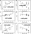
Non-cystic fibrosis bronchiectasis is increasingly described in the paediatric population. While diagnosis is by high-resolution chest computed tomography (CT), chest X-rays (CXRs) remain a first-line investigation. CXRs are currently insensitive in their detection of bronchiectasis. We aim to determine if quantitative digital analysis allows CT features of bronchiectasis to be detected in contemporaneously taken CXRs. Regions of radiologically (A) normal, (B) severe bronchiectasis, (C) mild airway dilation and (D) other parenchymal abnormalities were identified in CT and mapped to corresponding CXR. An artificial neural network (ANN) algorithm was used to characterise regions of classes A, B, C and D. The algorithm was then tested in 13 subjects and compared to CT scan features. Structural changes in CT were reflected in CXR, including mild airway dilation. The areas under the receiver operator curve for ANN feature detection were 0.74 (class A), 0.71 (class B), 0.76 (class C) and 0.86 (class D). CXR analysis identified CT measures of abnormality with a better correlation than standard radiological scoring at the 99% confidence level.Conclusion: Regional abnormalities can be detected by digital analysis of CXR, which may provide a low-cost and readily available tool to indicate the need for diagnostic CT and for ongoing disease monitoring. What is Known: • Bronchiectasis is a severe chronic respiratory disorder increasingly recognised in paediatric populations. • Diagnostic computed tomography imaging is often requested only after several chest X-ray investigations. What is New: • We show that a digital analysis of chest X-ray could provide more accurate identification of bronchiectasis features.
Keywords: Bronchiectasis; Chest X-rays; Children; Computed tomography; Image analysis.
© 2021. The Author(s), under exclusive licence to Springer-Verlag GmbH Germany, part of Springer Nature.
Conflict of interest statement
The authors declare no competing interests.
Figures


References
-
- Chang AB, Bell SC, Torzillo PJ, King PT, Maguire GP, Byrnes CA, Holland AE, O’Mara P, Grimwood K, group ev Chronic suppurative lung disease and bronchiectasis in children and adults in Australia and New Zealand Thoracic Society of Australia and New Zealand guidelines. Med J Aust. 2015;202:130. doi: 10.5694/mjac14.00287. - DOI - PubMed
-
- Singleton R, Valery P, Morris P, Byrnes C, Grimwood K, Redding G, Torzillo P, McCallum G, Chikoyak L, Mobberly C, Holman R, Chang A. Indigenous children from three countries with non-cystic fibrosis chronic suppurative lung disease/bronchiectasis. Pediatr Pulmonol. 2014;49:189–200. doi: 10.1002/ppul.22763. - DOI - PubMed
-
- Telfar B, Zhang J (2018) The impact of respiratory disease in New Zealand. 2018 update. Asthma and Respiratory Foundation of New Zealand, Wellington, New Zealand
MeSH terms
Grants and funding
LinkOut - more resources
Full Text Sources
Other Literature Sources

