Review
. 2021 Jun;22(3):215-235.
doi: 10.1007/s10162-021-00798-z. Epub 2021 Apr 28.

How Zebrafish Can Drive the Future of Genetic-based Hearing and Balance Research

Affiliations
Review

How Zebrafish Can Drive the Future of Genetic-based Hearing and Balance Research

Lavinia Sheets et al. J Assoc Res Otolaryngol. 2021 Jun.

Abstract

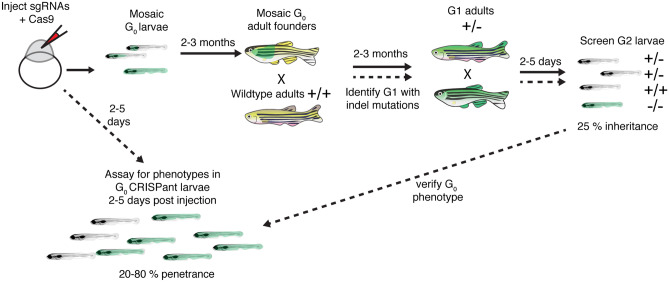
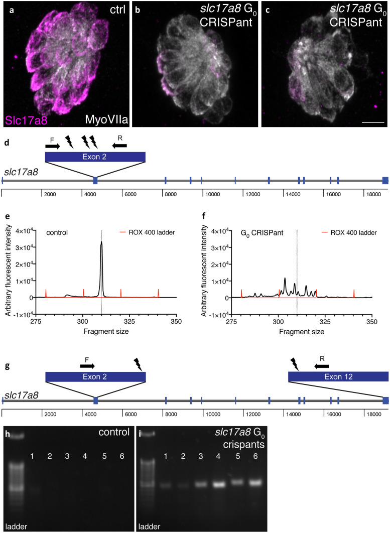
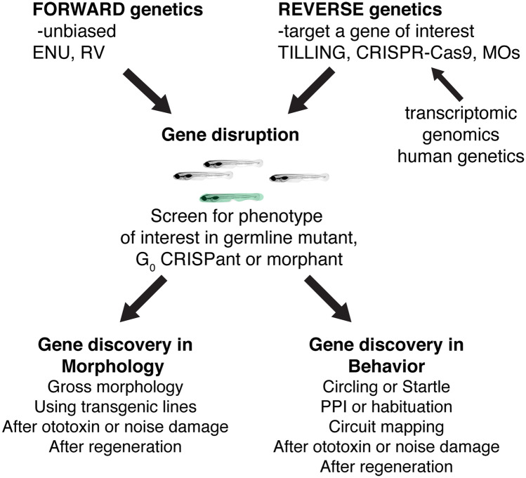
Over the last several decades, studies in humans and animal models have successfully identified numerous molecules required for hearing and balance. Many of these studies relied on unbiased forward genetic screens based on behavior or morphology to identify these molecules. Alongside forward genetic screens, reverse genetics has further driven the exploration of candidate molecules. This review provides an overview of the genetic studies that have established zebrafish as a genetic model for hearing and balance research. Further, we discuss how the unique advantages of zebrafish can be leveraged in future genetic studies. We explore strategies to design novel forward genetic screens based on morphological alterations using transgenic lines or behavioral changes following mechanical or acoustic damage. We also outline how recent advances in CRISPR-Cas9 can be applied to perform reverse genetic screens to validate large sequencing datasets. Overall, this review describes how future genetic studies in zebrafish can continue to advance our understanding of inherited and acquired hearing and balance disorders.

Keywords: genetic screening; genetics; hearing and balance; zebrafish.


Conflict of interest statement

The authors declare no competing interests.

Figures

Fig. 1
Fig. 1
Visualization of structures in hair cell systems of larval zebrafish. (a) A zebrafish larva at 5 days post fertilization (dpf) is shown. At this stage, both the inner ear and lateral line hair cell systems are functional. In this transgenic larvae, all hair cells are visualized via YFP fluorescence (Tg[myo6b:D3cpv]vo9 (Kindt et al. 2012)). (bb′) High magnification, side-view of hair cells in the medial crista (inner ear). A DIC image (b) and corresponding fluorescence image shows hair cells expressing β-actin-GFP to visualize hair bundles (b′, Tg(myo6b:actb1-EGFP)vo8 (Kindt et al. 2012)). (cc′) High magnification, top-down image of a neuromast hair cells expressing β-actin-GFP (c′, Tg(myo6b:actb1-EGFP)vo8) can also reveal hair bundle orientations (c′, arrows indicate orientations). (dd′) DIC image (d) and corresponding fluorescence image shows hair cells expressing Rib b-mCherry to label hair cell ribbons and afferent neurons expressing GFP to label the innervating fiber (d′, Tg(myo6b:ctbp2l-mCherry)idc3; Tg(neurod1:EGFP)nl1 (Sheets et al. ; Trapani et al. 2009). The image in a was taken at × 10, while all other images were taken at × 63 magnification. All images were taken at 5 dpf. Scale bar = 500 µm in a and 5 µm in b, c, and d
Fig. 2
Fig. 2
Outline of mutagenesis in zebrafish used for TILLING and forward genetic screening. Both retroviruses and the mutagen ENU can be used to create germline mutations in zebrafish. For retroviral-based mutagenesis, a DNA construct containing the retrovirus is injected into newly fertilized zebrafish embryos. These injected embryos are grown to adulthood (2–3 months), resulting in G0 adults that are mosaic for germline mutations. G0 adults are then outcrossed to wildtype adults to generate G1 adults that are heterozygous for different genetic lesions. In chemical mutagenesis, adult males are treated with a chemical mutagen such as ENU. These mutagenized males are crossed to wildtype adult females to generate G1 adults that are heterozygous for different genetic lesions. For a forward genetic screen (ENU or retroviral-based mutagens), G1 adults are crossed to wildtype animals, providing a pool of G2 adult carriers. G2 adults are incrossed, and G3 larvae are screened for a phenotype of interest. When mutagens are used to screen for a specific genetic lesion (in the case of TILLING), G1 adults or sperm stored in a library from G1 males are screened for mutations. After identifying a specific mutation, the identified G1 adult is crossed to wildtype to generate G2 adults harboring that mutation. Two G2 adults with the lesion of interest are then incrossed and screened for phenotypes. Blue and green represent distinct genetic lesions in mutagenized fish
Fig. 3
Fig. 3
GFP-based forward genetic screen reveals mutants with swollen afferent terminals. (ab) The Tg(neurod1:EGFP)nl1 transgenic line can be used to visualize the afferent terminals beneath hair cells in the lateral line (ab, green), while the vital dye FM 4-64 can be used to visualize lateral line hair cells (a, b, magenta). Actr10nl15 mutants were isolated in a forward genetic screen using Tg(neurod1:EGFP)nl1 to identify mutants with swollen lateral line afferent terminals (b) (Drerup et al. 2017). Hair cells in these mutant do not label with FM 4-64. Arrowheads indicate swellings in b. Images were taken at 5 dpf at × 63 magnification. Scale bar in b = 5 µm
Fig. 4
Fig. 4
Outline of Morpholino timeline in zebrafish. Morpholinos (MOs) are used to block mRNA splicing or translation of a particular gene product. MOs are injected into newly fertilized zebrafish embryos. Two to 5 days after MO injection morphant larvae can be screened for phenotype. The penetrance of MO phenotypes is highly variable. It is recommended that MO phenotypes be confirmed using a germline mutant when possible (Stainier et al. 2017)
Fig. 5
Fig. 5
Outline of how to create germline zebrafish CRISPR-Cas9 mutants and CRISPants. To create a germline zebrafish CRISPR-Cas9 mutant (follow path of solid lines), guide RNAs (gRNAs) targeting a gene of interest along with Cas9 mRNA or protein are injected into newly fertilized zebrafish embryos. These G0 injected embryos are grown to adulthood (2–3 months). G0 adult founders are crossed to wildtype adults, and the G1 progeny screened for indels. G1 progeny with indels are grown to adulthood. G1 adults containing indels are then incrossed and screened for phenotypes. To perform analyses on injected G0 CRISPant larvae, optimized gRNAs are injected into newly fertilized zebrafish embryos. Injected CRISPant G0 larvae are then screened for phenotypes days after the injection. CRISPant phenotypes can be verified by generating a stable, germline mutant
Fig. 6
Fig. 6
Two examples G0 slc17a8 CRISPant analysis and genotyping. Neuromasts from uninjected a and CRISPants embryos injected with the following gRNAs directed against the following sites in exon 2 of slc17a8 (5′-3′): GACAGAAGATGGTCGGCCGG (TGG), GGTGCTTTGGCCTTCCCAAA (CGG), and GCCCACCCCTATTGGACTGT (GGG) along with Cas9 protein bc. Staining with anti-Slc17a8 (Obholzer et al. 2008) and anti-MyosinVIIA (Developmental Studies Hybridoma Bank, #138-1) to label lateral line hair cells reveal that Slc17a8 staining is absent in G0 CRISPants that lack an acoustic startle response. Schematic of PCR analysis of slc17a8 d used to detect INDELs. The CRISPR-STAT assay, relying on fluorescent fragment analysis can be used to genotype individual CRISPants larvae and test gRNA efficiency. In these examples, there is a single peak in control larvae at 310 bp e. By comparison, in G0 slc17a8 CRISPants the peak at 310 bp is degraded, and numerous fragments (indicative of the many INDELs present in this mosaic founder) surrounding this peak are present f. Schematic of PCR analysis of slc17a8 g used to detect a large deletion. This PCR analysis was conducted on genomic DNA from uninjected control and CRISPant larvae lacking a startle response. Primers flank the sites targeted by the guides targeting exon 2 ((5′-3′)CACAGTCTACATCAACGGGA(CGG)) and exon 12 (TCCAGTGTAATGCACCATGG(AGG)) and were used to amplify the region between exon 2 and exon 12. Deletion of a 14.2-kb region in CRISPants yielded an ~ 400-bp PR product (lanes 1–6, i) that was absent in uninjected controls (lanes 1–6, h). Images in ac were taken at × 63 magnification on a Zeiss LSM 780 confocal microscope. Scale bar in c = 5 µm
Fig. 7
Fig. 7
Past, present, and future ways to use zebrafish genetics to study hearing and balance. Both forward and reverse genetic approaches in zebrafish have had an immense impact on gene discovery in hearing and balance. In the future, novel forward genetic screens using transgenic lines or novel damage paradigms have the potential to continue this path to gene discovery. In addition, advances in reverse genetics will continue to provide a valuable way to screen genes implicated in humans hearing loss. Reverse genetic screening many also prove a valuable, high-throughput pipeline to validate transcriptomics or genomics datasets

Similar articles

Cited by

References

    1. Abbas L, Whitfield TT. Nkcc1 (Slc12a2) is required for the regulation of endolymph volume in the otic vesicle and swim bladder volume in the zebrafish larva. Development. 2009;136:2837–2848. - PMC - PubMed
    1. Alexandre D, Ghysen A. Somatotopy of the lateral line projection in larval zebrafish. PNAS. 1999;96:7558–7562. - PMC - PubMed
    1. Almeida MP, Welker JM, Siddiqui S, Luiken J, Ekker SC, Clark KJ, Essner JJ, McGrail M. Endogenous zebrafish proneural Cre drivers generated by CRISPR/Cas9 short homology directed targeted integration. Sci Rep. 2021;11:1732. - PMC - PubMed
    1. Aman A, Nguyen M, Piotrowski T. Wnt/β-catenin dependent cell proliferation underlies segmented lateral line morphogenesis. Dev Biol. 2011;349:470–482. - PubMed
    1. Amsterdam A, Varshney G, Burgess S. Retroviral-mediated insertional mutagenesis in zebrafish. Methods Cell Biol. 2011;104:59–82. - PMC - PubMed

Publication types

LinkOut - more resources