. 2021 Oct;36(5):1052-1065.
doi: 10.1007/s12250-021-00368-w. Epub 2021 Apr 28.

A Virulent PEDV Strain FJzz1 with Genomic Mutations and Deletions at the High Passage Level Was Attenuated in Piglets via Serial Passage In Vitro

Affiliations

A Virulent PEDV Strain FJzz1 with Genomic Mutations and Deletions at the High Passage Level Was Attenuated in Piglets via Serial Passage In Vitro

Pengfei Chen et al. Virol Sin. 2021 Oct.

Abstract

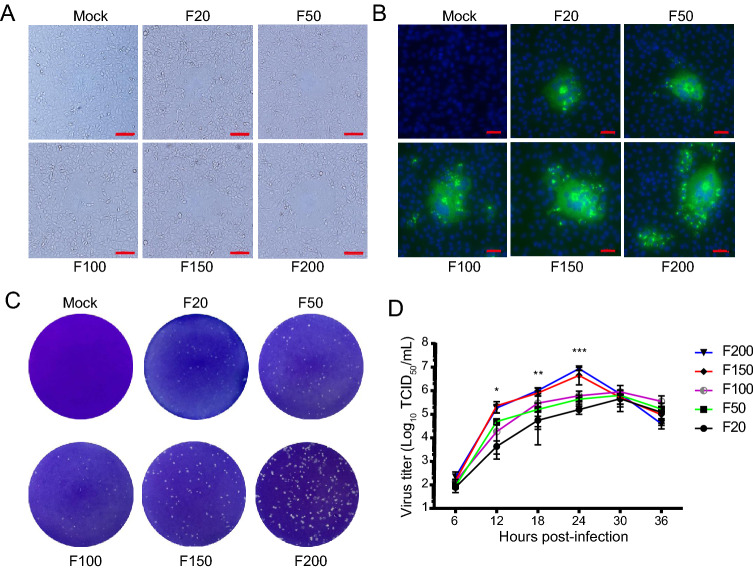
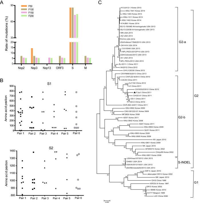
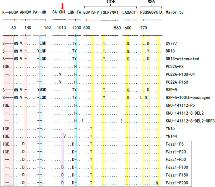
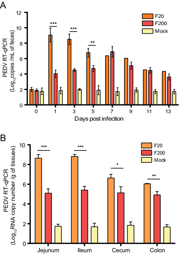
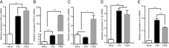
Highly virulent porcine epidemic diarrhea virus (PEDV) strains re-emerged and circulated in China at the end of 2010, causing significant economic losses in the pork industry worldwide. To understand the genetic dynamics of PEDV during its passage in vitro, the PEDV G2 strain FJzz1 was serially propagated in Vero cells for up to 200 passages. The susceptibility and adaptability of the FJzz1 strain increased gradually as it was serially passaged in vitro. Sequence analysis revealed that amino acid (aa) changes were mainly concentrated in the S glycoprotein, which accounted for 72.22%-85.71% of all aa changes. A continuous aa deletion (55I56G57E → 55K56Δ57Δ) occurred in the N-terminal domain of S1 (S1-NTD). To examine how the aa changes affected its virulence, FJzz1-F20 and FJzz1-F200 were selected to simultaneously evaluate their pathogenicity in suckling piglets. All the piglets in the FJzz1-F20-infected group showed typical diarrhea at 24 h postinfection, and the piglets died successively by 48 h postinfection. However, the clinical signs of the piglets in the FJzz1-F200-infected group were significantly weaker, and no deaths occurred. The FJzz1-F200-infected group also showed a lower level of fecal viral shedding and lower viral loads in the intestinal tissues, and no obvious histopathological lesions. Type I and III interferon were induced in the FJzz1-F200 infection group, together with pro-inflammatory cytokines, such as TNF-α, IL-1β and IL-8. These results indicate that the identified genetic changes may contribute to the attenuation of FJzz1 strain, and the attenuated FJzz1-F200 may have the potential for developing PEDV live-attenuated vaccines.

Keywords: Genetic variation; Pathogenicity; Porcine epidemic diarrhea virus (PEDV); Serially passage.


Conflict of interest statement

The authors declare that they have no conflict of interest.

Figures

Fig. 1
Fig. 1
Biological characterization of FJzz1 variants during serial passage in vitro. A Vero cells were mock infected or infected with FJzz1 variants F20, F50, F100, F150, or F200 at an MOI of 0.01, and a CPE was observed at 24 hpi. Scale bar = 100 µm. B Monolayers of Vero cells inoculated with FJzz1 variants F20, F50, F100, F150, or F200 were tested with an IFA using a MAb to PEDV N protein. Scale bar = 50 µm. C Crystal-violet-stained plaques formed on the monolayers of Vero cells inoculated with FJzz1 variants at different passages at 3 dpi. D Vero cells were infected with FJzz1 variants at different passages at an MOI of 0.01. The cell lysates were collected at the designated times and titrated with a TCID50 infectivity assay. Asterisk (*) indicates a significant difference between FJzz1-F20 and FJzz1-F200 (*P < 0.05; **P < 0.01; ***P < 0.001).
Fig. 2
Fig. 2
Phylogenetic analysis of FJzz1 strain. A Rate of mutation was calculated as the percentage of changed aa in the corresponding protein, including Nsp2, Nsp3, Nsp13, ORF3, S, and M. B Positions of the changed aa in the S protein are shown. The y-axis represents the positions of the changed aa, and the x-axis represents the six different pairs of virulent parental /attenuated strains (Pair 1: DR13/attenuated DR13; Pair 2: PC22A/PC22A-P160; Pair 3: 83P-5/83P-5-100th; Pair 4: KNU-141112-P5/KNU-141112-S-DEL2-ORF3; Pair 5: YN15/Y144; Pair 6: FJzz1-F20/FJzz1-F200). C A phylogenetic tree was constructed based on S gene of FJzz1 strain and another 60 reference strains with complete S gene sequences available in GenBank. The PEDV strain FJzz1 was marked with the filled triangle.
Fig. 3
Fig. 3
Alignment of S protein sequences of FJzz1 variants at different passages and other PEDV strains. S protein aa sequences of FJzz1 variants at different passages (marked with black box) and other PEDV strains including the classical CV777 strain, were aligned using Clustal W. Predicted N-linked glycosylation sites in the FJzz1 variants are marked with red arrows. Regions inserted and deleted relative to CV777 are highlighted in red and blue, respectively. The substitutions in neutralizing epitopes are highlighted in yellow.
Fig. 4
Fig. 4
Pathogenicity analysis of FJzz1-F20 and FJzz1-F200. A Fecal scores of piglets with the valuation standard: 0 = normal; 1 = soft; 2 = semi-fluid; 3 = watery diarrhea. B Changes in average body temperature in each group within the first 14 dpi. C Average bodyweight changes in each group. D Survival rates of piglets in each group. Survival curves of piglets infected with DMEM (Mock), FJzz1-F20, or FJzz1-F200 are shown.
Fig. 5
Fig. 5
Histopathological lesions in different intestinal segments of piglets inoculated with FJzz1-F20 or FJzz1-F200. A Different intestinal segments (Jejunum, Ileum, Cecum, and Colon) from FJzz1-F20-, FJzz1-F200- or mock-inoculated piglets were collected on the day of death or at the final time points for hematoxylin–eosin staining. B Immunohistochemical detection. Jejunal and Ileal tissues from each group were stained with a monoclonal antibody directed against PEDV N protein (1:200 dilution), scale bar = 200 μm.
Fig. 6
Fig. 6
Fecal viral shedding and quantification of viral load in different intestinal segments. Viral shedding in feces A and the viral loads in different intestinal segments B were determined with TaqMan real-time RT-PCR targeting the PEDV N gene. Asterisk (*) indicates a significant difference between FJzz1-F20 and FJzz1-F200 (*P < 0.05; **P < 0.01; ***P < 0.001).
Fig. 7
Fig. 7
Quantification of cytokines in jejunal tissue. Production of cytokines, including A type I interferon (IFN-α) and B type III interferon (IFN-λ3), and pro-inflammatory cytokines, such as C TNF-α, D IL-1β and E IL-8, were measured with real-time RT-PCR in jejunal tissue. The experiment was performed three times and the representative data are shown. Error bars represent means ± standard deviations (SD). *P < 0.05; **P < 0.01; ***P < 0.001.

References

    1. Chattha KS, Roth JA, Saif LJ. Strategies for design and application of enteric viral vaccines. Annu Rev Anim Biosci. 2015;3:375–395. - PubMed
    1. Chen F, Zhu Y, Wu M, Ku X, Ye S, Li Z, Guo X, He Q. Comparative genomic analysis of classical and variant virulent parental/attenuated strains of porcine epidemic diarrhea virus. Viruses. 2015;7:5525–5538. - PMC - PubMed
    1. Chen P, Wang K, Hou Y, Li H, Li X, Yu L, Jiang Y, Gao F, Tong W, Yu H, Yang Z, Tong G, Zhou Y. Genetic evolution analysis and pathogenicity assessment of porcine epidemic diarrhea virus strains circulating in part of China during 2011–2017. Infect Genet Evol. 2019;69:153–165. - PMC - PubMed
    1. Coussement W, Ducatelle R, Debouck P, Hoorens J. Pathology of experimental CV777 coronavirus enteritis in piglets. I Histological and histochemical study. Vet Pathol. 1982;19:46–56. - PubMed
    1. Crawford K, Lager KM, Kulshreshtha V, Miller LC, Faaberg KS. Status of vaccines for porcine epidemic diarrhea virus in the United States and Canada. Virus Res. 2016;226:108–116. - PubMed

LinkOut - more resources