Review
. 2021 Jul 1;321(1):C58-C71.
doi: 10.1152/ajpcell.00106.2021. Epub 2021 Apr 28.

Burn-induced hypermetabolism and skeletal muscle dysfunction

Affiliations
Review

Burn-induced hypermetabolism and skeletal muscle dysfunction

Carly M Knuth et al. Am J Physiol Cell Physiol. .

Abstract

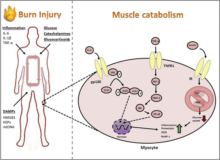
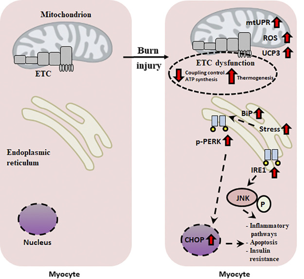
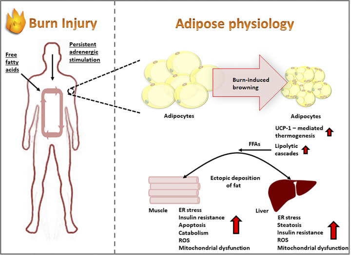
Critical illnesses, including sepsis, cancer cachexia, and burn injury, invoke a milieu of systemic metabolic and inflammatory derangements that ultimately results in increased energy expenditure leading to fat and lean mass catabolism. Burn injuries present a unique clinical challenge given the magnitude and duration of the hypermetabolic response compared with other forms of critical illness, which drastically increase the risk of morbidity and mortality. Skeletal muscle metabolism is particularly altered as a consequence of burn-induced hypermetabolism, as it primarily provides a main source of fuel in support of wound healing. Interestingly, muscle catabolism is sustained long after the wound has healed, indicating that additional mechanisms beyond wound healing are involved. In this review, we discuss the distinctive pathophysiological response to burn injury with a focus on skeletal muscle function and metabolism. We first examine the diverse consequences on skeletal muscle dysfunction between thermal, electrical, and chemical burns. We then provide a comprehensive overview of the known mechanisms underlying skeletal muscle dysfunction that may be attributed to hypermetabolism. Finally, we review the most promising current treatment options to mitigate muscle catabolism, and by extension improve morbidity and mortality, and end with future directions that have the potential to significantly improve patient care.

Keywords: inflammation; burn injury; hypermetabolism; muscle dysfunction.


Figures

Figure 1.
Figure 1.
Systemic factors released postburn induce skeletal muscle dysfunction. Proinflammatory cytokines such as interleukin-6 (IL-6), interleukin-1 β (IL-1β), and tumor necrosis factor α (TNF-α), as well as damage-associated molecular patterns (DAMPs) such as high-mobility group box 1 (HMGB1), heat shock proteins (HSPs), and mitochondrial DNA (mtDNA), induce muscle inflammation and proteolysis while perpetuating a state of insulin resistance. iNOS, inducible nitric oxide synthase; IR, insulin receptor; MuRF-1, muscle RING-finger protein-1.
Figure 2.
Figure 2.
Burn injury induces organellar dysfunction in muscle tissue. Mitochondrial uncoupling accompanied by increased ROS formation stimulates the unfolded protein response in both the mitochondria (mtUPR) and endoplasmic reticulum. Organellar stress contributes to apoptosis of muscle cells in addition to insulin resistance, a hallmark of burns. BiP, binding immunoglobulin protein; CHOP, C/EBP homologous protein; DAMP, damage-associated molecular pattern; ETC, electron transport chain; IRE1, inositol-requiring enzyme 1; JNK, c-Jun N-terminal kinase; PERK, protein kinase R (PKR)-like endoplasmic reticulum kinase; UCP3, uncoupling protein-3; UPR, unfolded protein response; ROS, reactive oxygen species.
Figure 3.
Figure 3.
Dysfunctional organ cross talk as a product of severe burn injuries. A severe burn induces the pathological browning of subcutaneous white adipose tissue, forming thermogenic “beige” fat, which is highly lipolytic. In turn, adipose modulates the function of downstream tissues such as the liver and muscle via the release of FFAs, perpetuating systemic dysfunction postburn. ER, endoplasmic reticulum; FFAs, free fatty acids; ROS, reactive oxygen species; UCP1, uncoupling protein-1.

Similar articles

Cited by

References

    1. Mortality GBD and Causes of Death Collaborators. Global, regional, and national age-sex specific all-cause and cause-specific mortality for 240 causes of death, 1990-2013: a systematic analysis for the Global Burden of Disease Study 2013. Lancet 385: 117–171, 2015. doi:10.1016/S0140-6736(14)61682-2. - DOI - PMC - PubMed
    1. Brooks NC, Marshall AH, Qa’aty N, Hiyama Y, Boehning D, Jeschke MG. XBP-1s is linked to suppressed gluconeogenesis in the Ebb phase of burn injury. Mol Med 19: 72–78, 2013. doi:10.2119/molmed.2012.00348. - DOI - PMC - PubMed
    1. Sommerhalder C, Blears E, Murton AJ, Porter C, Finnerty C, Herndon DN. Current problems in burn hypermetabolism. Curr Probl Surg 57: 100709, 2020. doi:10.1016/j.cpsurg.2019.100709. - DOI - PMC - PubMed
    1. Jeschke MG, Gauglitz GG, Kulp GA, Finnerty CC, Williams FN, Kraft R, Suman OE, Mlcak RP, Herndon DN. Long-term persistence of the pathophysiologic response to severe burn injury. PLoS One 6: e21245, 2011. doi:10.1371/journal.pone.0021245. - DOI - PMC - PubMed
    1. Stanojcic M, Abdullahi A, Rehou S, Parousis A, Jeschke MG. Pathophysiological response to burn injury in adults. Ann Surg 267: 576–584, 2018. doi:10.1097/SLA.0000000000002097. - DOI - PMC - PubMed

Publication types

MeSH terms