. 2021 Apr 28;19(1):86.
doi: 10.1186/s12915-021-01017-0.

Rat PRDM9 shapes recombination landscapes, duration of meiosis, gametogenesis, and age of fertility

Affiliations

Rat PRDM9 shapes recombination landscapes, duration of meiosis, gametogenesis, and age of fertility

Ondrej Mihola et al. BMC Biol. .

Abstract

Background: Vertebrate meiotic recombination events are concentrated in regions (hotspots) that display open chromatin marks, such as trimethylation of lysines 4 and 36 of histone 3 (H3K4me3 and H3K36me3). Mouse and human PRDM9 proteins catalyze H3K4me3 and H3K36me3 and determine hotspot positions, whereas other vertebrates lacking PRDM9 recombine in regions with chromatin already opened for another function, such as gene promoters. While these other vertebrate species lacking PRDM9 remain fertile, inactivation of the mouse Prdm9 gene, which shifts the hotspots to the functional regions (including promoters), typically causes gross fertility reduction; and the reasons for these species differences are not clear.

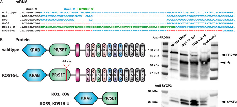
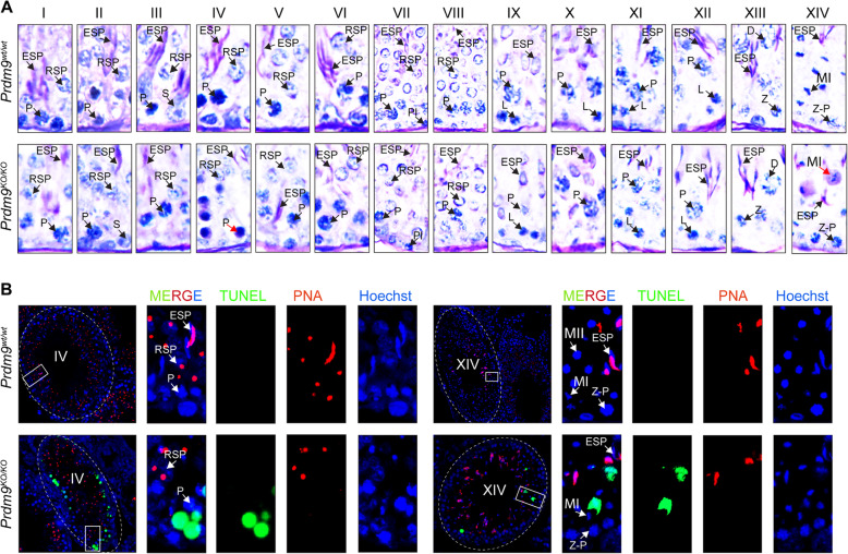
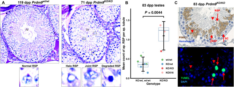
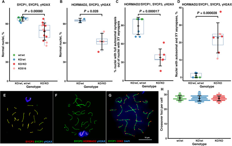
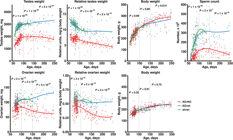
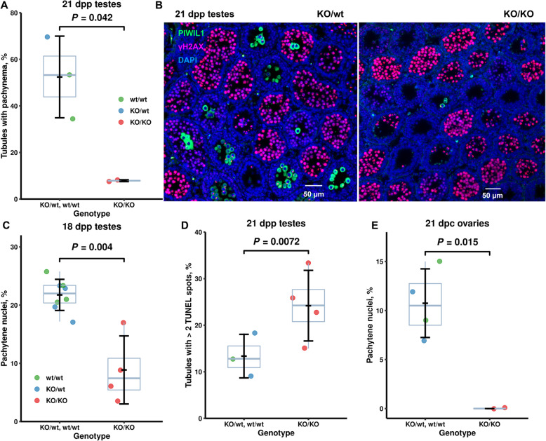
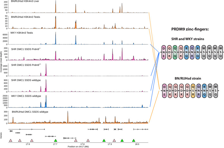
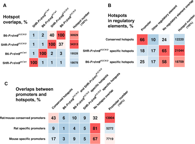
Results: We introduced Prdm9 deletions into the Rattus norvegicus genome and generated the first rat genome-wide maps of recombination-initiating double-strand break hotspots. Rat strains carrying the same wild-type Prdm9 allele shared 88% hotspots but strains with different Prdm9 alleles only 3%. After Prdm9 deletion, rat hotspots relocated to functional regions, about 40% to positions corresponding to Prdm9-independent mouse hotspots, including promoters. Despite the hotspot relocation and decreased fertility, Prdm9-deficient rats of the SHR/OlaIpcv strain produced healthy offspring. The percentage of normal pachytene spermatocytes in SHR-Prdm9 mutants was almost double than in the PWD male mouse oligospermic sterile mutants. We previously found a correlation between the crossover rate and sperm presence in mouse Prdm9 mutants. The crossover rate of SHR is more similar to sperm-carrying mutant mice, but it did not fully explain the fertility of the SHR mutants. Besides mild meiotic arrests at rat tubular stages IV (mid-pachytene) and XIV (metaphase), we also detected postmeiotic apoptosis of round spermatids. We found delayed meiosis and age-dependent fertility in both sexes of the SHR mutants.

Conclusions: We hypothesize that the relative increased fertility of rat versus mouse Prdm9 mutants could be ascribed to extended duration of meiotic prophase I. While rat PRDM9 shapes meiotic recombination landscapes, it is unnecessary for recombination. We suggest that PRDM9 has additional roles in spermatogenesis and speciation-spermatid development and reproductive age-that may help to explain male-specific hybrid sterility.

Keywords: Fertility; Meiotic recombination; PRDM9; Rattus norvegicus.


Conflict of interest statement

The authors declare that they have no competing interests.

Figures

Fig. 1
Fig. 1
Four Prdm9 deletions generated in SHR rats lead to mRNAs with open reading frame truncations and one also to alternative mRNA with deletion of 20 codons. a mRNAs arising from rats with genomic deletions of 2-, 8-, 39-, and 516-bp (abbreviated KO2, KO8, KO39, and KO516, respectively; the latter two also involve a part of Intron 8); blue dashes, gaps to optimize the alignment; red dashes, exonic deletions; -U, -L, two new mRNAs from the animal with the KO516 deletion. b Polypeptide products predicted from mRNAs in a and their detection. -20 a.a., deletion of 20 amino acid residues. The C-terminal boxes indicate zinc-fingers and the three letters their DNA-binding amino acids. See Additional file 1: Fig. S1 for details of the translations and amino acids essential for methyltransferase activity and Additional file 2: Table S1 for genomic and cDNA sequences. Right, PRDM9 was detected at the expected sizes of 97 kDa (mouse Dom2 allele) and 96 kDa (rat SHR allele). Anti-SYCP3 antibody was utilized as a loading control. PRDM9 was present in the SHR-Prdm9KO516/KO516 mutants at the expected size of the KO516-L isoform (91 kDa), but its expression was lower than that of the wild-type in SHR; the KO516-U isoform (predicted 40 kDa) was undetectable. KO39 mutant product was not found (expected 44 kDa) in the SHR-Prdm9KO39/KO39 testes, thus no C-terminally truncated form of PRDM9 was seen in either mutant. Only a third of the amount of protein nuclear extract was loaded in mouse (10 μg) compared to rat (30 μg) lanes
Fig. 2
Fig. 2
Decreased fertility parameters of SHR males carrying Prdm9 deletions depend on age. Each dot depicts a single animal. a–c Boxplots of sperm count (a), testicular weight (b), and relative testicular weight (c, mg testicular weight per gram of body weight). d, Representative images of hematoxylin-eosin (HE) stained sections from rat testicles; note the presence of mature spermatids (arrows) in all three males. e Litter size of male parents of two indicated Prdm9 genotypes for paternal age at birth. The statistical significance of the differences between genotypes was tested using LRM in the age groups of up to 150 and above 150 dpp. f Number of pups per month per female (OMU) for mating ranges (days) in SHR males of two indicated Prdm9 genotypes. The data underlying all published plots are given in Additional file 3
Fig. 3
Fig. 3
Decreased fertility and premature ovarian failure in SHR females carrying Prdm9 deletions. a, b Boxplots of ovary weight (a, in mg) and of relative ovary weight (b, mg ovary weight per gram of body weight). c Examples of HE stained sections from three adult rat ovaries at two magnifications. Note that all females carry germ cell follicles (arrows). d Litter size of female parents of two indicated Prdm9 genotypes with maternal age at birth. The difference between genotypes was tested using the LRM in the age group of up to 150 dpp. e Number of pups per month per female (OMU) and mating ranges (days) for two indicated Prdm9 genotypes. The abscissa at OMU = 0 contains data for 14 Prdm9-deficient females, ten of which were mated after 150 dpp (for additional mean of 26 days). f Follicle quantification from ovarian sections of the indicated Prdm9 genotypes at three points of postnatal development. Differences between three Prdm9 genotypes and between animals of three ages within each genotype were analyzed using LRM with subsequent Tukey test and Bonferroni adjustment. g Representative images of HE-stained ovarian sections from 21-dpp females
Fig. 4
Fig. 4
Prdm9-deficiency causes spermatogenic arrests in tubular stages IV and XIV of SHR rat. a Testicular histology using PAS-H staining. Illustrative examples of all 14 rat tubule stages. Degenerated cells in seminiferous tubules from adult (71 dpp) mutants stain intensively with PAS-H (blue-purple) at stages IV and XIV (red arrows). P, pachytene spermatocytes; RSP, round spermatids; ESP, elongated spermatids; S, Sertoli cells; Pl, preleptotene; L, leptotene; D, diplotene; Z-P, zygotene-pachytene spermatocytes; MI, metaphase I spermatocytes. Quantification of cells in all stages is presented in Fig. 5. b Increased apoptosis in mutant rat stage IV and XIV tubules confirmed by labeling apoptosis (TUNEL), acrosome (PNA), and DNA (Hoechst 33342)
Fig. 5
Fig. 5
Prdm9-deficiency changes the quantities of spermatogenic cells in all 14 tubular stages of SHR rat. Cell counts of spermatogenic cell types in seminiferous tubules of stages I to XIV are given including the mean cell number ± standard deviation per tubule in each stage calculated from 3 to 5 tubules for each stage and animal (4 mutants and 3 controls) from sections stained with PAS-H. SG A, type A spermatogonia; SG In, intermediated spermatogonia; SG B, type B spermatogonia; see legend of Fig. 4 for abbreviations of primary spermatocytes and spermatids; MII, metaphase II spermatocytes; sSC, secondary spermatocytes; dM, degenerated MI/II and sSC; dP, degenerated pachytene; dSP, degenerated, halo, and joint spermatids
Fig. 6
Fig. 6
Rat PRDM9 affects postmeiotic development of round spermatids. a Examples of normal and defective spermatids from wild-type and Prdm9-deficient SHR males on PAS-H stained testicular sections. The means counted in mutant versus control in all 14 stages are in Fig. 5. b, c Increased apoptosis during postmeiotic development of Prdm9-deficient SHR testes. b Mean counts of apoptotic round spermatids per apoptotic tubule were scored from randomly chosen apoptotic tubules (18 to 32 per animal, total 221 tubules from 9 animals), plotted, and evaluated using LRM. The average counts of mutant and control samples were 1.1 ± 0.4 and 0.4 ± 0.2, respectively. c Round spermatids (RSP) were distinguished on neighboring TUNEL-DAPI and anti-PIWIL1-Hematoxylin-stained sections from spermatocytes (SC); representative images (the same tubule) are shown
Fig. 7
Fig. 7
Decreased homologous synapsis but similar crossover rate in SHR males with Prdm9 deletions compared to wild-type. a, b Percentage of normal pachytene spermatocytes (with all autosomes synapsed). Each dot represents a single animal (over 50 cells). Antibodies used for the staining of chromosomal spreads are given in the headings of each graph. Nuclear spreads used for the analysis of autosomal synapsis in both a and b were used for the analysis of XY synapsis in c and d. c Percentage of nuclei with full autosomal synapsis out of nuclei with XY asynapsis in Prdm9-deficient and control rat testes. d percentage of nuclei with both autosomal and XY asynapsis in Prdm9-deficient and control rat testes. e, f, Examples of normal pachytene cells. Immunocytochemistry with antibodies against: e Synaptonemal complex (SYCP1, SYCP3) and chromatin surrounding DSB sites (γH2AX); f SYCP3, γH2AX, and unsynapsed chromosomal axes (HORMAD2). See Additional file 1: Fig. S6 for the images of representative nuclei from Prdm9-deficient males. Differences between Prdm9 genotypes were analyzed using LRM with subsequent Tukey test and Holm adjustment for multiple testing. g Representative image of chromosomal spread from a Prdm9-deficient rat immunostained for cyclin-dependent kinase 2 (CDK2) and synaptonemal complex (SYCP1), confirming that CDK2 localizes both to crossover nodules and telomeres as in the mouse. h Counts of autosomal crossover nodules per cell (N = 2 animals for both Prdm9KO/wt and Prdm9KO/KO, N = 1 for wild-type)
Fig. 8
Fig. 8
Fertility parameters of Prdm9-deficient animals change with age. Differences between three Prdm9 genotypes were analyzed using LRM with subsequent Tukey tests and Holm adjustment in three age groups: 50–100, 100–150, and 150–250 dpp. P values illustrate differences between SHR-Prdm9KO/wt and SHR-Prdm9KO/KO rats. The comparison of wild-type and SHR-Prdm9KO/wt rats revealed no differences in fertility parameters and had highly consistent correlation curves. The correlation between age and body weight was used as a negative control
Fig. 9
Fig. 9
Delayed meiosis in male and female SHR rats lacking Prdm9 function. a-d Delayed prophase I in Prdm9-deficient male rats compared to controls. a, b Immunohistochemistry of 21-dpp testes (154 to 368 tubules scored from each animal). c Immunocytochemistry of 18-dpp testicular spreads (over 400 spermatocytes for each genotype). d Delayed meiosis is associated with apoptosis. Immunohistochemistry of prepubertal testes using TUNEL and DAPI. e Delayed onset of pachynema in Prdm9-deficient female rats. Spread 21-dpc ovarian nuclei were staged using immunocytochemistry (over 250 oocytes scored for each of the three Prdm9 genotypes; LRM). The data underlying all published plots are in Additional file 3
Fig. 10
Fig. 10
The positions of the rat meiotic DSB hotspots are determined by PRDM9. Left, a region of rat Chromosome 17 (rn5 assembly) exemplifying that the DSB hotspots (shown as peaks) in wild-type SHR males (SHR Prdm9+/+ SSDS) are shared with the strain with the same Prdm9 allele (WKY Prdm9+/+ SSDS), but not with the strain with different Prdm9 allele (BN/RIJHsd Prdm9+/+ SSDS) or inactivated Prdm9 allele (SHR Prdm9−/− SSDS). DSB hotspots in Prdm9-deficient male co-localize with testicular (T) and less with liver (L) H3K4me3 marks. The H3K4me3 profiles from the BN a WKY strains are different and reflect hotspot sites unique to each of these strains, thus validating that rat PRDM9 displays H3-methyltransferase activity. Some promoters correspond to Prdm9-independent hotspots (green arrowheads), but other promoters (pink arrowheads) are not targeted. The row “Genes” shows Ensembl gene models. Raw ChIP-seq coverage is shown in 150-bp windows; each panel is scaled to the maximum value. Right, DNA-binding zinc-finger arrays of PRDM9 from two rat strains showing polymorphic amino acid residues in contact with DNA
Fig. 11
Fig. 11
Overlaps of recombination initiation hotspots from Prdm9-deficient rodents with a wild-type hotspots and b regulatory elements. c Overlaps of promoters with hotspots

References

    1. Bolcun-Filas E, Schimenti JC. Genetics of meiosis and recombination in mice. Int Rev Cell Mol Biol. 2012;298:179–227. doi: 10.1016/B978-0-12-394309-5.00005-5. - DOI - PubMed
    1. Baudat F, Buard J, Grey C, Fledel-Alon A, Ober C, Przeworski M, Coop G, de Massy B. PRDM9 is a major determinant of meiotic recombination hotspots in humans and mice. Science. 2010;327(5967):836–840. doi: 10.1126/science.1183439. - DOI - PMC - PubMed
    1. Myers S, Bowden R, Tumian A, Bontrop RE, Freeman C, MacFie TS, McVean G, Donnelly P. Drive against hotspot motifs in primates implicates the PRDM9 gene in meiotic recombination. Science. 2010;327(5967):876–879. doi: 10.1126/science.1182363. - DOI - PMC - PubMed
    1. Parvanov ED, Petkov PM, Paigen K. Prdm9 controls activation of mammalian recombination hotspots. Science. 2010;327(5967):835. doi: 10.1126/science.1181495. - DOI - PMC - PubMed
    1. Pratto F, Brick K, Khil P, Smagulova F, Petukhova GV, Camerini-Otero RD. DNA recombination. Recombination initiation maps of individual human genomes Science. 2014;346(6211):1256442. - PMC - PubMed

Publication types