Novel clinical presentation of a CRX rod-cone dystrophy
- PMID: 33910785
- PMCID: PMC8094365
- DOI: 10.1136/bcr-2019-233711
Novel clinical presentation of a CRX rod-cone dystrophy
Abstract
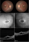
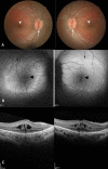
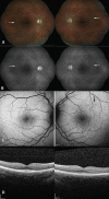
We describe a novel clinical presentation of a CRX rod-cone dystrophy in a single family. Two boys ages 6 and 12 years presented with clinical and optical coherence tomography features suggestive of X-linked retinoschisis, but with optic nerve swelling without increased intracranial pressure. One patient had an electronegative electroretinogram (ERG) and the other had rod-cone dysfunction. Neither had retinoschisin (RS1) gene mutations. Biological mother and sister presented with retinal pigment epithelium (RPE) changes and abnormal cone-rod ERG responses. On further testing, next generation sequencing with array comparative genomic hybridisation showed a deletion in exon 4 of the CRX gene. Cystoid maculopathy in young male children can be difficult to distinguish from RS1-associated schisis. Phenotypic variants within a family must prompt a thorough retinal dystrophy evaluation even with electronegative ERG in the presenting child. This novel phenotype for CRX presents with optic nerve swelling and cystoid maculopathy in men, and RPE changes in women.
Keywords: genetics; macula; ophthalmology; retina.
© BMJ Publishing Group Limited 2021. No commercial re-use. See rights and permissions. Published by BMJ.
Conflict of interest statement
Competing interests: None declared.
Figures


Similar articles
-
A recurrent arcuate retinopathy in familial cone-rod dystrophy secondary to heterozygous CRX deletion.Ophthalmic Genet. 2019 Dec;40(6):493-499. doi: 10.1080/13816810.2019.1688841. Epub 2019 Nov 19. Ophthalmic Genet. 2019. PMID: 31743059
-
A novel gene mutation in a family with X-linked retinoschisis.J Formos Med Assoc. 2015 Sep;114(9):872-80. doi: 10.1016/j.jfma.2014.01.001. Epub 2014 Feb 13. J Formos Med Assoc. 2015. PMID: 24529551
-
Childhood cone-rod dystrophy with macular cystic degeneration from recessive CRB1 mutation.Ophthalmic Genet. 2014 Sep;35(3):130-7. doi: 10.3109/13816810.2013.804097. Epub 2013 Jun 14. Ophthalmic Genet. 2014. PMID: 23767994
-
Of men and mice: Human X-linked retinoschisis and fidelity in mouse modeling.Prog Retin Eye Res. 2022 Mar;87:100999. doi: 10.1016/j.preteyeres.2021.100999. Epub 2021 Aug 11. Prog Retin Eye Res. 2022. PMID: 34390869 Review.
-
The Value of Electroretinography in Identifying Candidate Genes for Inherited Retinal Dystrophies: A Diagnostic Guide.Diagnostics (Basel). 2023 Sep 25;13(19):3041. doi: 10.3390/diagnostics13193041. Diagnostics (Basel). 2023. PMID: 37835784 Free PMC article. Review.
Cited by
-
Cone Rod Homeobox (CRX): literature review and new insights.Ophthalmic Genet. 2025 Aug;46(4):338-346. doi: 10.1080/13816810.2025.2458086. Epub 2025 Mar 12. Ophthalmic Genet. 2025. PMID: 40074530 Review.
-
Gene Augmentation for Autosomal Dominant CRX-Associated Retinopathies.Adv Exp Med Biol. 2023;1415:135-141. doi: 10.1007/978-3-031-27681-1_21. Adv Exp Med Biol. 2023. PMID: 37440026 Free PMC article. Review.
References
Publication types
MeSH terms
LinkOut - more resources
Full Text Sources
Other Literature Sources
Medical
