

APOSTEL 2.0 Recommendations for Reporting Quantitative Optical Coherence Tomography Studies
- PMID: 33910937
- PMCID: PMC8279566
- DOI: 10.1212/WNL.0000000000012125
APOSTEL 2.0 Recommendations for Reporting Quantitative Optical Coherence Tomography Studies
Abstract
Objective: To update the consensus recommendations for reporting of quantitative optical coherence tomography (OCT) study results, thus revising the previously published Advised Protocol for OCT Study Terminology and Elements (APOSTEL) recommendations.
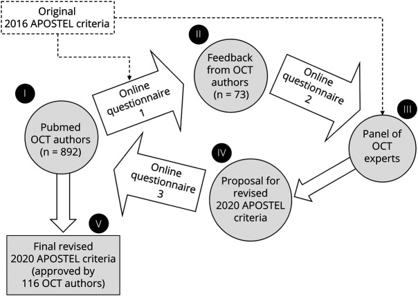
Methods: To identify studies reporting quantitative OCT results, we performed a PubMed search for the terms "quantitative" and "optical coherence tomography" from 2015 to 2017. Corresponding authors of the identified publications were invited to provide feedback on the initial APOSTEL recommendations via online surveys following the principle of a modified Delphi method. The results were evaluated and discussed by a panel of experts and changes to the initial recommendations were proposed. A final survey was recirculated among the corresponding authors to obtain a majority vote on the proposed changes.
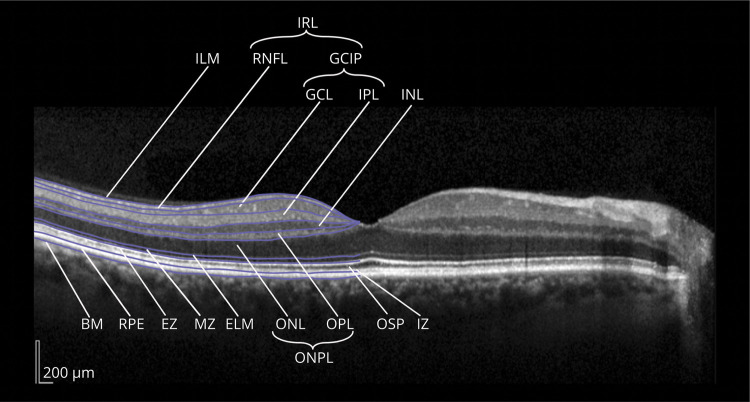
Results: A total of 116 authors participated in the surveys, resulting in 15 suggestions, of which 12 were finally accepted and incorporated into an updated 9-point checklist. We harmonized the nomenclature of the outer retinal layers, added the exact area of measurement to the description of volume scans, and suggested reporting device-specific features. We advised to address potential bias in manual segmentation or manual correction of segmentation errors. References to specific reporting guidelines and room light conditions were removed. The participants' consensus with the recommendations increased from 80% for the previous APOSTEL version to greater than 90%.
Conclusions: The modified Delphi method resulted in an expert-led guideline (evidence Class III; Grading of Recommendations, Assessment, Development and Evaluations [GRADE] criteria) concerning study protocol, acquisition device, acquisition settings, scanning protocol, funduscopic imaging, postacquisition data selection, postacquisition analysis, nomenclature and abbreviations, and statistical approach. It will be essential to update these recommendations to new research and practices regularly.
Copyright © 2021 The Author(s). Published by Wolters Kluwer Health, Inc. on behalf of the American Academy of Neurology.
Figures
References
-
- Guyatt GH, Oxman AD, Schünemann HJ, Tugwell P, Knottnerus A. GRADE guidelines: a new series of articles in the Journal of Clinical Epidemiology. J Clin Epidemiol. 2011;64(4):380-382. - PubMed
-
- de Villiers MR, de Villiers PJ, Kent AP. The Delphi technique in health sciences education research. Med Teach. 2005;27(7):639-643. - PubMed
-
- Cameron JR, Albrecht P, Cruz-Herranz A, Petzold A, Lagreze WA, Brandt AU. The APOSTEL recommendations for reporting quantitative optical coherence tomography studies. Neurology. 2016;87(24):1960. - PubMed
MeSH terms
Grants and funding
LinkOut - more resources
Full Text Sources
Other Literature Sources
Medical