. 2021 Apr 28;13(591):eabe7378.
doi: 10.1126/scitranslmed.abe7378.

SynNotch-CAR T cells overcome challenges of specificity, heterogeneity, and persistence in treating glioblastoma

Affiliations

SynNotch-CAR T cells overcome challenges of specificity, heterogeneity, and persistence in treating glioblastoma

Joseph H Choe et al. Sci Transl Med. .

Abstract

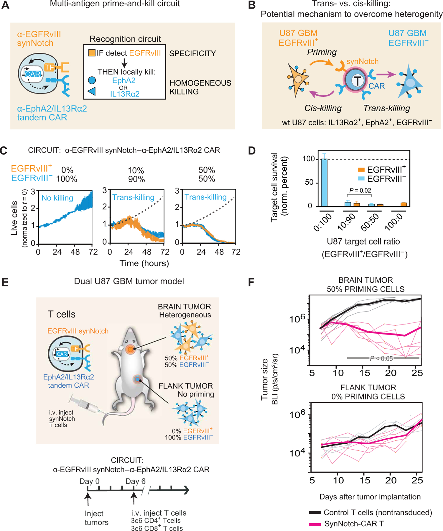
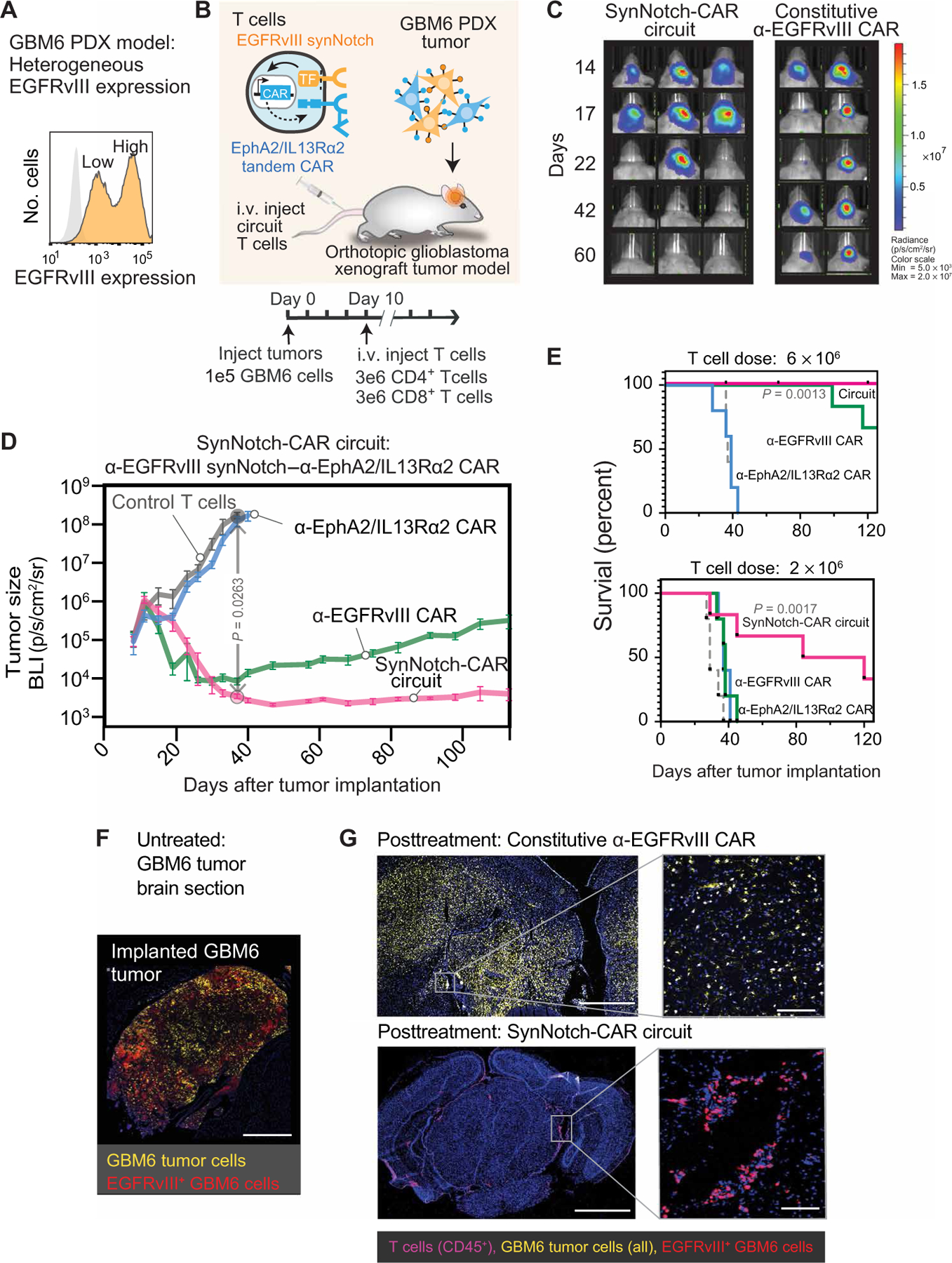
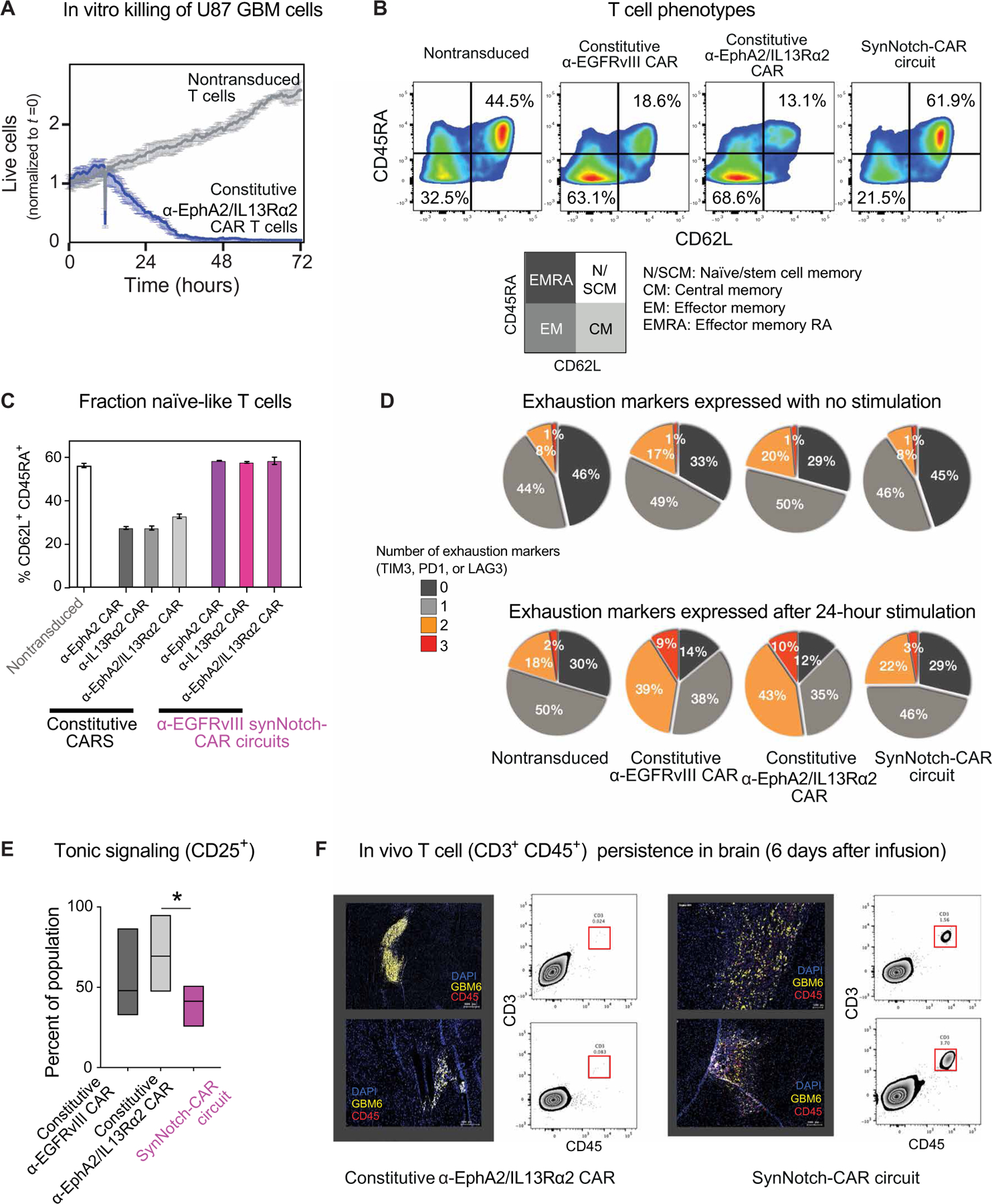
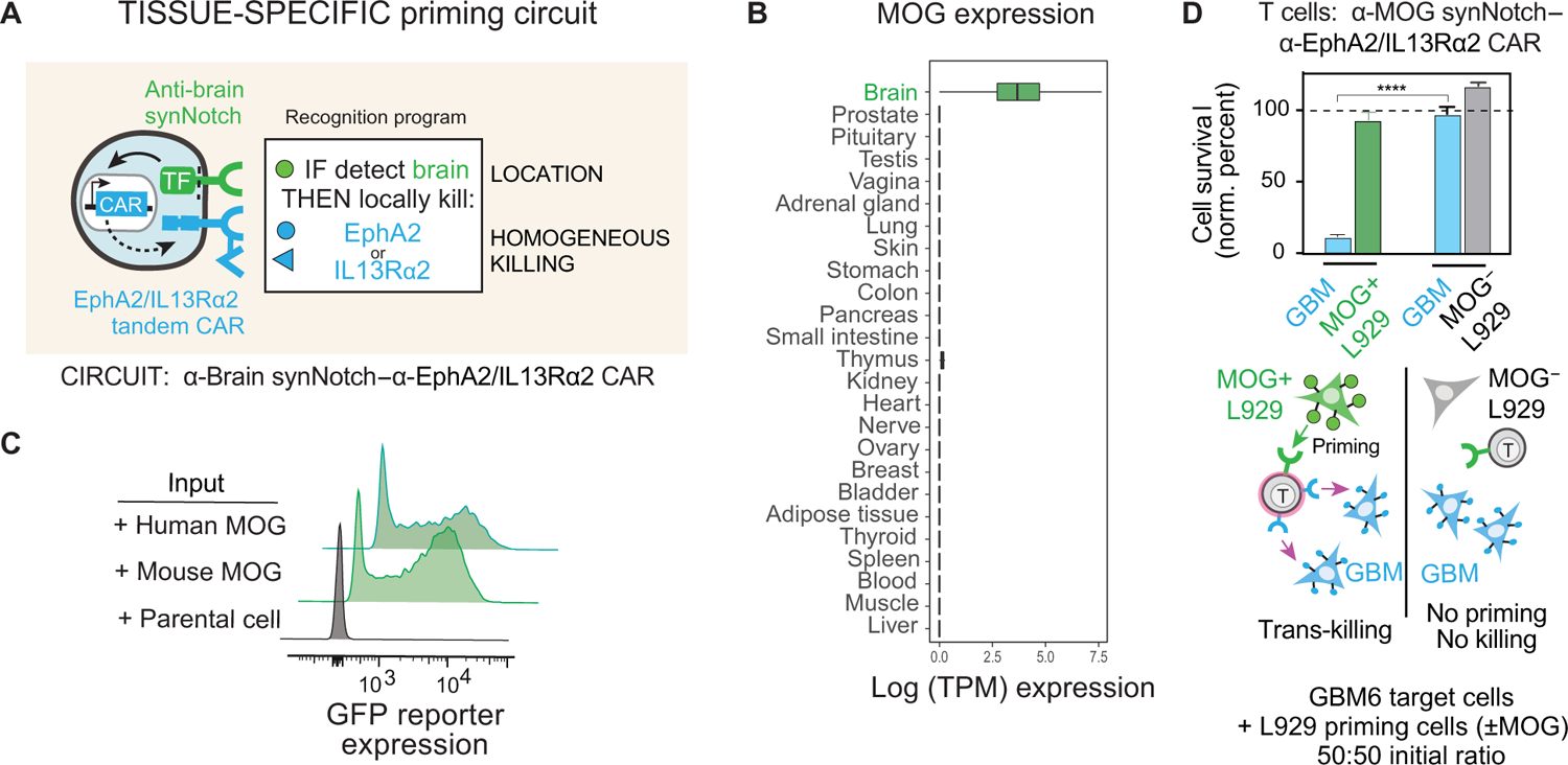
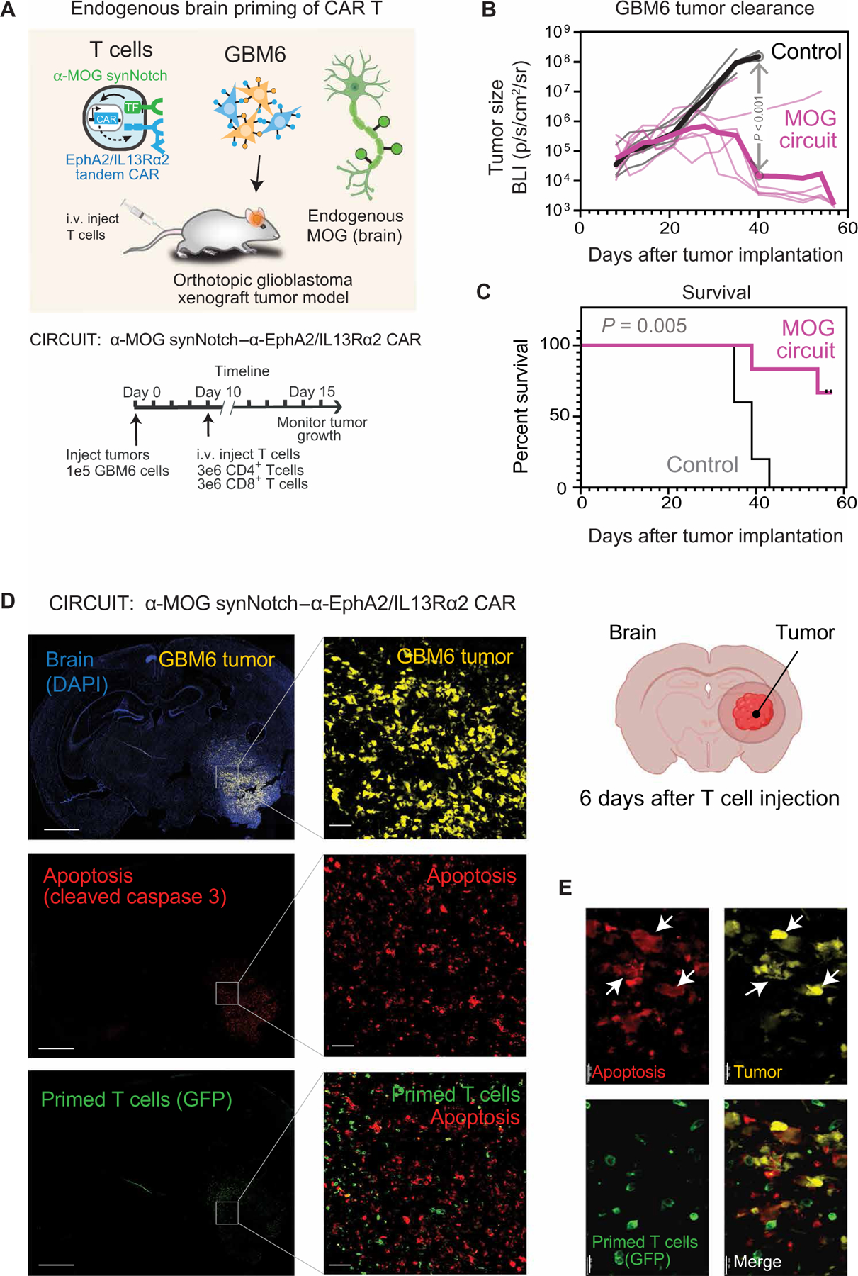
Treatment of solid cancers with chimeric antigen receptor (CAR) T cells is plagued by the lack of ideal target antigens that are both absolutely tumor specific and homogeneously expressed. We show that multi-antigen prime-and-kill recognition circuits provide flexibility and precision to overcome these challenges in the context of glioblastoma. A synNotch receptor that recognizes a specific priming antigen, such as the heterogeneous but tumor-specific glioblastoma neoantigen epidermal growth factor receptor splice variant III (EGFRvIII) or the central nervous system (CNS) tissue-specific antigen myelin oligodendrocyte glycoprotein (MOG), can be used to locally induce expression of a CAR. This enables thorough but controlled tumor cell killing by targeting antigens that are homogeneous but not absolutely tumor specific. Moreover, synNotch-regulated CAR expression averts tonic signaling and exhaustion, maintaining a higher fraction of the T cells in a naïve/stem cell memory state. In immunodeficient mice bearing intracerebral patient-derived xenografts (PDXs) with heterogeneous expression of EGFRvIII, a single intravenous infusion of EGFRvIII synNotch-CAR T cells demonstrated higher antitumor efficacy and T cell durability than conventional constitutively expressed CAR T cells, without off-tumor killing. T cells transduced with a synNotch-CAR circuit primed by the CNS-specific antigen MOG also exhibited precise and potent control of intracerebral PDX without evidence of priming outside of the brain. In summary, by using circuits that integrate recognition of multiple imperfect but complementary antigens, we improve the specificity, completeness, and persistence of T cells directed against glioblastoma, providing a general recognition strategy applicable to other solid tumors.


Conflict of interest statement

Competing interests: W.A.L. is on the Scientific Advisory Board for Allogene Therapeutics and is a shareholder of Gilead Sciences and Intellia Therapeutics. H.O. is on the Scientific Advisory Board for Neuvogen and Eureka Therapeutics. K.T.R. is a cofounder of Arsenal Biosciences. W.A.L., H.O., P.B.W., and J.H.C. are inventors on the following relevant patent/patent applications held/submitted by UCSF: Trans-antigen targeting in heterogeneous cancers and methods of use thereof (WO 2019/195576 A1) (2019); Methods of treating EGFRvIII-expressing glioblastomas (WO2019/195586 A1; US 62/654,012) (2019); Methods of treating glioblastomas (WO2019195596A1) (2019); W.A.L., H.O., P.B.W., J.H.C., and M.S.S. are inventors on the following relevant patent/patent applications held/submitted by UCSF: Use of MOG for priming a treatment for glioblastoma (US 62/980,882) (2020) and Use of brain-specific antigens to home, block and deliver cell-based treatments to the brain (US 62/980,885) (2020). H.O. is an inventor on patent Treatment of cancer using humanized anti-EGFRvIII chimeric antigen receptor US20190330356A1 (2019) held by University of Pennsylvania and University of Pittsburgh that covers EGFRvIII targeting.

Figures

Fig. 1.
Fig. 1.. Multi-antigen prime-and-kill circuits in T cells provide a general strategy to overcome antigen heterogeneity while still maintaining high tumor specificity.
(A) Design of synNotch-CAR circuit primed by EGFRvIII neoantigen: α-EGFRvIII synNotch receptor induces expression of tandem α-EphA2/IL13Rα2 CAR (TF, transcription factor). These cells should be activated to kill EphA2+ or IL13Rα2+ target cells only if exposed to EGFRvIII+ cells. (B) Such T cells could overcome priming antigen heterogeneity if they can execute trans-killing, where priming and killing antigens are expressed on different but neighboring cells. (C) Real-time killing assays using heterogeneous mixtures of EGFRvIII+ and EGFRvIII target cells show efficient trans-killing. Primary CD8+ human T cells transduced with α-EGFRvIII synNotch–α-EphA2/IL13Rα2 CAR circuit were cultured with indicated ratios of EGFRvIII+ versus EGFRVIII U87 cells at an E:T ratio of 5:1 and imaged over 3 days using IncuCyte. The EGFRvIII+ cell population (priming cells) is shown in yellow, and the EGFRvIII cell population (target cells) is shown in blue. The presence of as low as 10% priming cells yielded strong killing of EGFRvIII target cells, although killing was slightly slower compared to that observed with 50% priming cells (P = 0.0149 and P = 0.0218, t test at 48 and 72 hours, respectively). The dotted black line shows the growth of 100% target cells as a reference (n = 3, error bars denote SEM). See movie S1. (D) Relative survival of each cell type in experiments from (C) (at 72 hours). Killing of target cells is efficiently primed by as low as 10% priming cells (EGFRvIII+). No killing is observed in the absence of priming cells (n = 3, error bars denote SD). A t test was used for statistical comparison. (E) NCG mice were simultaneously implanted with two GBM tumors: a heterogeneous tumor comprising EGFRvIII+ and EGFRvIII U87 cells (1:1 ratio) in the brain, and a homogeneous EGFRvIII U87 tumor implanted subcutaneously in the flank. Mice were treated 6 days after tumor implantation with intravenous (i.v.) infusion of 3 million CD4+ and CD8+ synNotch-CAR T cells (n = 6) or control nontransduced T cells (n = 6). (F) Tumor size was measured by luciferase bioluminescence imaging (BLI) over time as the number of photons per second per square centimeter per steradian (p/s/cm2/sr). Tumor size curves for individual mice treated with synNotch-CAR T cells are shown in light pink; curves for mice treated with nontransduced T cells are shown in gray. Thicker lines correspond to geometric means. P < 0.05 by Mann-Whitney test on day 12 and onward, whereas the flank tumor grew at the same rate as in the mice treated with nontransduced T cells.
Fig. 2.
Fig. 2.. SynNotch-CAR T cells show improved efficacy and durability compared to individual parental constitutive CARs in clearing heterogeneous GBM6 PDX tumors.
(A) Flow cytometry analysis of a patient-derived xenograft (PDX) GBM6 tumor model shows intrinsic heterogeneity of EGFRvIII expression. (B) Timeline for in vivo tumor experiments with GBM6 tumors. GBM6 tumors expressing mCherry and luciferase were orthotopically implanted in brains of NCG mice. Ten days after tumor implantation, mice were infused intravenously (i.v.) with 3 million each of CD4+ and CD8+ T cells expressing no construct (control) (n = 5), α-EGFRvIII synNotch–α-EphA2/IL13Rα2 CAR circuit (n = 6), constitutively expressed α-EGFRvIII CAR (n = 5), or constitutively expressed α-EphA2/IL13Rα2 tandem CAR (n = 5). (C) Longitudinal bioluminescence imaging of GBM6 tumor–bearing mice treated with α-EGFRvIII synNotch–α-EphA2/IL13Rα2 CAR T cells and conventional α-EGFRvIII CAR T cells. Each column represents one mouse over time. (D) Time course of tumor size measured by bioluminescence. P = 0.0263, two-way ANOVA followed by a Dunnett’s test nontransduced versus synNotch-CAR T cells at day 37. Error bars represent means ± SEM of five to six individual mice from one experiment. (E) Kaplan-Meier survival curves for high-dose (6 × 10⁶ cells) and low-dose (2 × 10⁶ cells) treatments. Statistical significance was calculated using log-rank Mantel-Cox test. (F) Fluorescence microscopy of representative section of untreated GBM6 xenograft tumor, isolated 15 days after tumor implantation, shows heterogeneous expression of EGFRvIII. Scale bar, 500 µm. (G) Top: Representative fluorescence microscopy of a brain and tumor section isolated 107 days after treatment with conventional α-EGFRvIII CAR T cells shows the presence of tumor but loss of EGFRvIII expression. Bottom: Representative fluorescence microscopy of a brain section isolated 110 days after treatment with EGFRvIII synNotch–α-EphA2/IL13Rα2 CAR T cells reveals clearance of a GBM6 xenograft tumor and sustained presence of T cells. Scale bars, 1 mm (left) and 50 µm (right).
Fig. 3.
Fig. 3.. Early time point imaging shows thatA priming and killing of synNotch-CAR T cells are precisely restricted to tumor region.
(A) Representative confocal fluorescent microscopy of brain slices from GBM6 tumor–bearing mice isolated at 6 days after infusion with α-EGFRvIII synNotch–α-EphA2/IL13Rα2 CAR T cells reveals primed T cells expressing CAR-GFP only within the tumor bed. Cleaved caspase 3 is also only observed within the tumor. Scale bars, 1 mm (left) and 100 µm (right). Overlay of tumor, cleaved caspase 3, and primed GFP T cell is shown in fig. S5B. (B) Representative confocal images of brain slices from GBM6 tumor–bearing mice isolated at 6 days after treatment with nontransduced T cells. Scale bar, 1 mm. (C) Intravital two-photon imaging of synNotch-CAR T cells shows priming within the GBM6 xenograft tumor. Tumors were implanted at a depth of 3 mm below the right frontal cortex, and cranial windows were implanted. Scale bar, 400 µm. See movie S2. (D) Half-life of CAR expression decay in synNotch-CAR T cells after removing priming cells. T cells were primed in vitro with GBM6 cells for 60 hours and then isolated by cell sorting. GFP-tagged CAR expression was observed over time. MFI, median fluorescence intensity. Decay experiment was performed in the absence (top) or presence (bottom) of target U87 cells lacking priming antigen. Exponential decay fit was used to determine the CAR half-life. Decay curves depict one representative of four different donors.
Fig. 4.
Fig. 4.. SynNotch-CAR T cells show enhanced naïve/stem cell memory phenotype, reduced exhaustion, and improved persistence in vivo.
(A) In vitro killing by T cells constitutively expressing α-EphA2/IL13Rα2 tandem CAR (relative to nontransduced control). (B) Flow cytometry plots distinguishing naïve-like cells (CD45RA+CD62L+), central memory cells (CD45RACD62L+), effector memory cells (CD45RACD62L), and effector memory RA cells (CD45RA+CD62L) (representative of three experiments from different donors). Percentages of cells in the naïve/memory stem cell state and effector memory state are highlighted. T cells were rested for 10 days in vitro after transfection before phenotypic analysis. (C) Percentage of CD62L+CD45RA+ T cells in indicated CAR T cells and synNotch-CAR T cells (n = 3 per group). (D) Expression of exhaustion markers by indicated CAR and synNotch-CAR T cells, both without and with stimulation by target cells. Pie chart shows percentage of cells that express 0, 1, 2, or 3 exhaustion markers (PD1, LAG3, or TIM3), average of three different donors. (E) Tonic signaling in constitutive versus α-EGFRvIII-synNotch induced α-EphA2/IL13Rα2 CAR T cells, measured by percent CD25+ cells (one-way ANOVA followed by a Dunnett’s test, P < 0.05, n = 5 different donors). Boxes represent min to max with median center line. (F) SynNotch-CAR T cells show improved persistence in vivo compared to constitutive CAR T cells. GBM6 tumor–bearing mice were euthanized 6 days after infusion of either the constitutive tandem CAR T cells (left) or α-EGFRvIII synNotch–α-EphA2/IL13Rα2 CAR T cells (right). Representative confocal fluorescent microscopy of brain slices and single-cell suspensions of tumor-bearing brain show few T cells (CD3+CD45+) in constitutive CAR T cell–treated tumors. In contrast, mice treated with synNotch-CAR T cells reveal high number of T cells in tumors.
Fig. 5.
Fig. 5.. Design of brain antigen-primed synNotch-CAR circuit to target GBM.
(A) “Tissue-specific priming” circuit is designed to restrict killing only to the brain, preventing damage to normal, nonbrain tissues that express killing antigens, EphA2 and IL13Rα2. In principle, this circuit should selectively identify GBM cells as the only brain-localized cells that are EphA2+ or IL13Rα2+. (B) Box and whisker plots showing tissue-specific expression of MOG across a subset of tissue samples in GTEx v7. Units shown are log scale–normalized RNA-sequencing counts (transcripts per million) taken from GTEx portal v7 (https://gtexportal.org/). (C) Primary CD8+ T cells expressing α-MOG synNotch and GFP reporter were cocultured with either parental K562 cells or K562 cells expressing mouse or human MOG. Flow cytometry histograms show induction of GFP reporter only in the presence of MOG+ K562 cells (representative of three experiments). (D) Primary CD8+ T cells transduced with α-MOG synNotch–α-EphA2/IL13Rα2 CAR circuit were cocultured with GBM6 target cells and L929 priming cells either expressing or not expressing mouse MOG. Relative cell survival of both target GBM and priming L929 cells was tracked over 72 hours (n = 3, error bars indicate SD). ****P < 0.0001, t test compared to those cocultured with MOG L929 cells. The cell population ratio was 1:1:1, 1 × 10⁴ cells each. No significant killing of the L929 priming cell population was observed in either experiment.
Fig. 6.
Fig. 6.. Tissue-specific priming of synNotch-CAR T cells by brain-specific antigen MOG induces effective killing of GBM6 brain tumors in vivo.
(A) GBM6 tumor cells were stereotactically implanted into brains of NCG mice. GBM6 cells were engineered to express mCherry and luciferase to allow for tracking of tumor size. Ten days after tumor implantation, mice were infused intravenously with 3 million each of CD4+ and CD8+ T cells. T cells expressed either no construct (nontransduced control) (n = 5) or α-MOG synNotch–α-EphA2/IL13Rα2 CAR circuit (n = 6). (B and C) Tumor size (B) and survival (C) were monitored over time. Tumor size was determined by bioluminescence imaging. Negative control treatment (nontransduced T cells) is shown in gray, and α-MOG SynNotch-CAR circuit treatment is shown in pink. P < 0.001 by t test with Holm-Sidak correction for multiple comparisons (on day 40). Thin lines show traces for individual animals; thick line shows geometric mean. See fig. S9G for analysis of off-target specificity (using non–brain-implanted tumor). Survival was analyzed over 60 days by log-rank (Mantel-Cox) test. P = 0.005. (D) GBM6 tumor–bearing mice were euthanized 6 days after α-MOG SynNotch-CAR T cell infusion. Representative confocal fluorescent microscopy of brain sections reveals that T cell–mediated killing (cleaved caspase 3 staining) is restricted to the tumor. Scale bars, 2 mm (left) and 50 µm (right). (E) Insets (single-stained images) are enlargements of outlined regions in main images. Expression of cleaved caspase 3 is confined to the tumor cells (yellow), as indicated by white arrows in the overlay image. Scale bar, 20 µm.

Comment in

References

    1. June CH, Sadelain M, Chimeric antigen receptor therapy. N. Engl. J. Med 379, 64–73 (2018). - PMC - PubMed
    1. Johnson LA, Morgan RA, Dudley ME, Cassard L, Yang JC, Hughes MS, Kammula US, Royal RE, Sherry RM, Wunderlich JR, Lee C-CR, Restifo NP, Schwarz SL, Cogdill AP, Bishop RJ, Kim H, Brewer CC, Rudy SF, VanWaes C, Davis JL, Mathur A, Ripley RT, Nathan DA, Laurencot CM, Rosenberg SA, Gene therapy with human and mouse T-cell receptors mediates cancer regression and targets normal tissues expressing cognate antigen. Blood 114, 535–546 (2009). - PMC - PubMed
    1. Parkhurst MR, Yang JC, Langan RC, Dudley ME, Nathan D-AN, Feldman SA, Davis JL, Morgan RA, Merino MJ, Sherry RM, Hughes MS, Kammula US, Phan GQ, Lim RM, Wank SA, Restifo NP, Robbins PF, Laurencot CM, Rosenberg SA, T cells targeting carcinoembryonic antigen can mediate regression of metastatic colorectal cancer but induce severe transient colitis. Mol. Ther. J. Am. Soc. Gene Ther 19, 620–626 (2011). - PMC - PubMed
    1. Morgan RA, Chinnasamy N, Abate-Daga D, Gros A, Robbins PF, Zheng Z, Dudley ME, Feldman SA, Yang JC, Sherry RM, Phan GQ, Hughes MS, Kammula US, Miller AD, Hessman CJ, Stewart AA, Restifo NP, Quezado MM, Alimchandani M, Rosenberg AZ, Nath A, Wang T, Bielekova B, Wuest SC, Akula N, McMahon FJ, Wilde S, Mosetter B, Schendel DJ, Laurencot CM, Rosenberg SA, Cancer regression and neurological toxicity following anti-MAGE-A3 TCR gene therapy. J. Immunother 36, 133–151 (2013). - PMC - PubMed
    1. Morgan RA, Yang JC, Kitano M, Dudley ME, Laurencot CM, Rosenberg SA, Case report of a serious adverse event following the administration of T cells transduced with a chimeric antigen receptor recognizing ERBB2. Mol. Ther. J. Am. Soc. Gene Ther 18, 843–851 (2010). - PMC - PubMed

Publication types

Substances