Review
. 2021 Apr 28;85(2):e00031-20.
doi: 10.1128/MMBR.00031-20. Print 2021 May 19.

Incompatibility Group I1 (IncI1) Plasmids: Their Genetics, Biology, and Public Health Relevance

Affiliations
Review

Incompatibility Group I1 (IncI1) Plasmids: Their Genetics, Biology, and Public Health Relevance

Steven L Foley et al. Microbiol Mol Biol Rev. .

Abstract

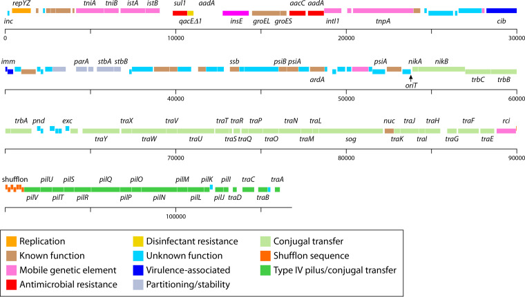
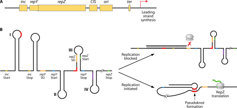
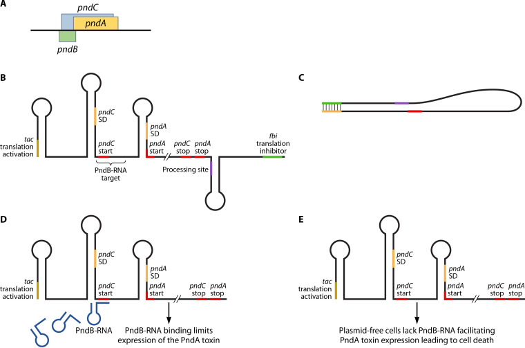
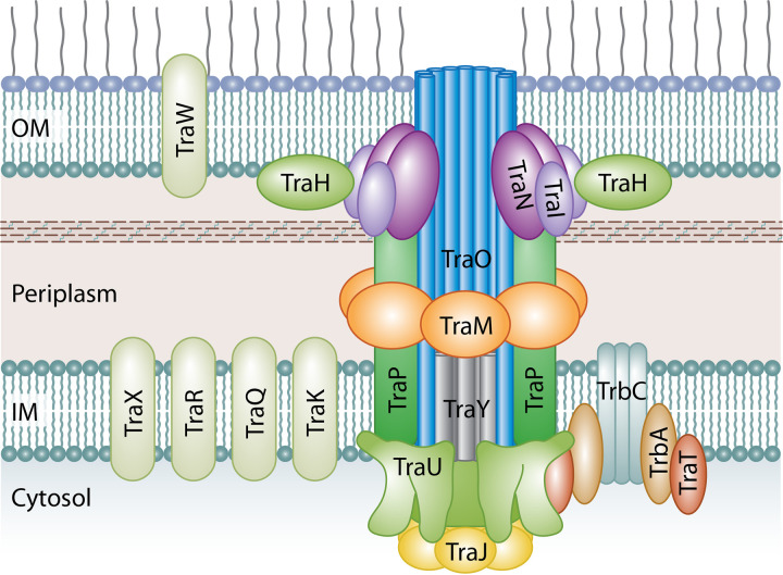
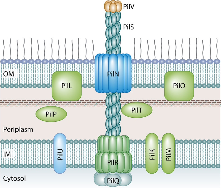
Bacterial plasmids are extrachromosomal genetic elements that often carry antimicrobial resistance (AMR) genes and genes encoding increased virulence and can be transmissible among bacteria by conjugation. One key group of plasmids is the incompatibility group I1 (IncI1) plasmids, which have been isolated from multiple Enterobacteriaceae of food animal origin and clinically ill human patients. The IncI group of plasmids were initially characterized due to their sensitivity to the filamentous bacteriophage If1. Two prototypical IncI1 plasmids, R64 and pColIb-P9, have been extensively studied, and the plasmids consist of unique regions associated with plasmid replication, plasmid stability/maintenance, transfer machinery apparatus, single-stranded DNA transfer, and antimicrobial resistance. IncI1 plasmids are somewhat unique in that they encode two types of sex pili, a thick, rigid pilus necessary for mating and a thin, flexible pilus that helps stabilize bacteria for plasmid transfer in liquid environments. A key public health concern with IncI1 plasmids is their ability to carry antimicrobial resistance genes, including those associated with critically important antimicrobials used to treat severe cases of enteric infections, including the third-generation cephalosporins. Because of the potential importance of these plasmids, this review focuses on the distribution of the plasmids, their phenotypic characteristics associated with antimicrobial resistance and virulence, and their replication, maintenance, and transfer.

Keywords: antimicrobial resistance; incompatibility group I1 plasmids; plasmid biology; plasmid genetics; plasmid maintenance; plasmid replication; plasmid transfer; public health; virulence.


Figures

FIG 1
FIG 1
Overview of the major regions that generally make up IncI1-type plasmids as described by Sampei et al. (25). These regions include the plasmid replication and control regions, variable regions encoding antimicrobial resistance and/or virulence-associated genes, genes associated with plasmid stability and partitioning, the leading region, which may play a role in conjugal transfer, and regions associated with conjugal transfer (25). The plasmid represented is pSH1148_107, GenBank accession number JN983049 (43).
FIG 2
FIG 2
Genetic map of IncI1 plasmid pSH1148_107, GenBank accession number JN983049 (43). The gene names of known genes are included along with color coding of the predicted functions of each of the genes (25).
FIG 3
FIG 3
(A) Diagram of the replication control region for IncI1 plasmids. RepZ is the main replication initiation protein and interacts with the origin of replication (ori), which is near repZ, to initiate replication of the plasmid sequence. Termination of plasmid replication occurs at CIS, which is located between repZ and ori (57). (B) Predicted RNA structure of the replication control (Rep) region of the IncI1 plasmid and predicted mechanisms of replication control. Control of repZ translation, and subsequently control of plasmid replication and copy number, is associated with the negative regulator inc and the positive regulator repY. To control replication, inc mRNA binds to the inc sequence and blocks the ribosomal binding site to inhibit RepY translation. To activate replication, inc mRNA is unbound from inc, allowing translation of RepY, which facilitates pseudoknot formation (binding of structure I to structure III at the binding sites indicated in red) that opens the ribosomal binding site to facilitate RepZ expression (based on data from reference 55).
FIG 4
FIG 4
Diagram of the Pnd toxin-antitoxin system. The system can encode the PndA toxin that causes lethal damage to the bacterial cell membrane when expressed. (A) The Pnd operon is made up of pndC that overlaps pndA in the plasmid sequence, and pndB is located on the opposite DNA strand. pndC modulates pndA expression, and pndB encodes an RNA molecule that suppresses expression of pndA and toxin formation. (B) Transcription of the pnd operon leads to the formation of a complex mRNA molecule whose translation is regulated by multiple mechanisms. The 5′ end of the inactive RNA molecule contains a translational activation element (tac), and the 3′ end contains a fold-back inhibition element (fbi). Between these elements are the nucleotides for the translation of PndC and PndA and a processing site for the formation of the functional mRNA molecules. (C) The fbi and tac sites are complementary to one another and bind to prevent translation of the unprocessed RNA molecule. (D) Following processing (cleavage at the processing site and removal of the fbi element), the regulation of the translation of the Pnd toxin in the activated mRNA is due to the binding of the small, very short-lived PndB RNA molecules that bind to the more stable PndCA mRNA overlapping the translation start site for PndC. (E) In cases where the plasmids are lost, all of the short-lived PndB are degraded, allowing the translation of the long-lived PndA toxin leading to cell membrane damage and cell death.
FIG 5
FIG 5
Predicted structure of the Tra/Trb T4SS of IncI1 plasmids based on homologs from the better-characterized Dot/Icm and Ti T4SSs. The inner membrane portion of the T4SS is predicted to be made up of TraJ, TraM, TraO, TraP, TraU, and TraY proteins. TraP forms a multimer with TraO and TraY through the inner membrane and into the periplasmic space where the complex appears to interact with the outer membrane complex. TraU has homology to DotO of the Dot/Icm T4SS, which forms a hexamer that sits at the base of the main pore channel of the secretion system where it interacts with the TraJ multimer. TraJ homologs are predicted to form hexamers that are on the cytoplasmic side of the secretion system and form a transitory complex with the TraU multimer in line with the core complex. TrbC is a T4CP that forms a complex with TrbA and TraT and functions to help deliver macromolecules from the cytoplasm to the T4SS section apparatus to traverse the cell membranes. The outer membrane complex made up of 13 subunits comprised of TraI, TraH, and TraN surrounds the TraO secretion channel consisting of 18 subunits. OM, outer membrane; IM, inner membrane.
FIG 6
FIG 6
Predicted structure of the T4P of IncI1 plasmids based on homologs from the better-characterized TCP and BFG pili. PilS forms the major prepilin polymer complex that extends from the cell to form the T4P and is capped by PilV subunits that make up the minor prepilin multimer that interacts with specific oligosaccharide in LPS on a recipient cell. Other structural elements include PilR, which is the integral inner membrane spanning protein, and PilN, which forms the outer membrane secretin through which the PilS polymer extends. PilT is predicted to be a lytic transglycosidase that may function to create a pore through the peptidoglycan layer to allow elongation of the pilus structure. PilR proteins are predicted to be platform proteins that transfer energy from the system ATPases, such as PilQ, to the T4P structure.

Similar articles

Cited by

References

    1. Johnson TJ, Nolan LK. 2009. Plasmid replicon typing. Methods Mol Biol 551:27–35. 10.1007/978-1-60327-999-4_3. - DOI - PubMed
    1. Gualberto JM, Kuhn K. 2014. DNA-binding proteins in plant mitochondria: implications for transcription. Mitochondrion 19(Pt B):323–328. 10.1016/j.mito.2014.02.004. - DOI - PubMed
    1. Johnson TJ, Lang KS. 2012. IncA/C plasmids: an emerging threat to human and animal health? Mob Genet Elements 2:55–58. 10.4161/mge.19626. - DOI - PMC - PubMed
    1. Johnson TJ, Nolan LK. 2009. Pathogenomics of the virulence plasmids of Escherichia coli. Microbiol Mol Biol Rev 73:750–774. 10.1128/MMBR.00015-09. - DOI - PMC - PubMed
    1. Frost LS, Koraimann G. 2010. Regulation of bacterial conjugation: balancing opportunity with adversity. Future Microbiol 5:1057–1071. 10.2217/fmb.10.70. - DOI - PubMed

Publication types

LinkOut - more resources