. 2021 Apr 28;11(1):9219.
doi: 10.1038/s41598-021-88615-8.

MicroRNA-138 suppresses glioblastoma proliferation through downregulation of CD44

Affiliations

MicroRNA-138 suppresses glioblastoma proliferation through downregulation of CD44

Margaret Yeh et al. Sci Rep. .

Abstract

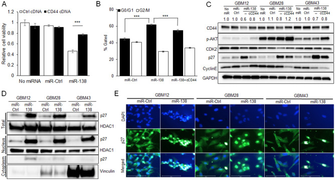
Tumor suppressive microRNAs (miRNAs) are increasingly implicated in the development of anti-tumor therapy by reprogramming gene network that are aberrantly regulated in cancer cells. This study aimed to determine the therapeutic potential of putative tumor suppressive miRNA, miR-138, against glioblastoma (GBM). Whole transcriptome and miRNA expression profiling analyses on human GBM patient tissues identified miR-138 as one of the significantly downregulated miRNAs with an inverse correlation with CD44 expression. Transient overexpression of miR-138 in GBM cells inhibited cell proliferation, cell cycle, migration, and wound healing capability. We unveiled that miR-138 negatively regulates the expression of CD44 by directly binding to the 3' UTR of CD44. CD44 inhibition by miR-138 resulted in an inhibition of glioblastoma cell proliferation in vitro through cell cycle arrest as evidenced by a significant induction of p27 and its translocation into nucleus. Ectopic expression of miR-138 also increased survival rates in mice that had an intracranial xenograft tumor derived from human patient-derived primary GBM cells. In conclusion, we demonstrated a therapeutic potential of tumor suppressive miR-138 through direct downregulation of CD44 for the treatment of primary GBM.


Conflict of interest statement

The authors declare no competing interests.

Figures

Figure 1
Figure 1
Downregulation of miR-138 in glioblastoma (GBM) patient specimens. (A) Principal component analyses (PCA) on miRNA expression profiling obtained by NanoString miRNA Expression Analysis from 9 GBM patients and 4 negative control samples. As a quality control, PCA plot clearly classified the samples into two groups. (B) Volcano plot reveals down-regulation of miR-138 in an inverse correlation with miR-21. (C) Expression of miR-138 in GBM samples (0.009915 ± 0.003592, n = 9) compared to negative control samples (0.3525 ± 0.08532, n = 4) analyzed by qRT-PCR using TaqMan individual miRNA assay. (D) The Cancer Genome Atlas Program (TCGA) database shows that miR-138 expression is downregulated in glioblastoma (GBM) compared to normal tissues. All error bars indicates standard deviations, and Student t-test was used to determine the significance in difference between the two groups. ***p < 0.001.
Figure 2
Figure 2
Overexpression of miR-138 inhibits cell proliferation and migration in vitro. (A) GBM cell proliferation with miR-138 overexpression. Patient-derived primary GBM cells (GBM12, 28 and 43) were transfected with a series of concentration of miR-138 mimics or negative control (miR-Ctrl). After 4 days of transfection, viable cells were measured by CellTiter-Glo Luminescent Cell Viability Assay. (B) Cell proliferation analysis by fluorescence live cell imaging every four hours on GBM12-RFP cells after transfection of 25 nM miR-138 or miR-Ctrl. (C) Representative fluorescence images of GBM12-RFP cells at 96 h after transfection of 25 nM miR-138 or miR-Ctrl. (D) Apoptosis analysis on GBM cells (GBM12, 28 and 43) after transfection of 25 nM miR-138 or miR-Ctrl followed by Annexin V-PI double staining for flow cytometry. Percentage of apoptotic cell populations were indicated in red-dotted box on each representative cytograms (n = 3). (E) Cell migration assay in Trans-well chamber plates. GBM cells transfected with 25 nM miR-138 or miR-Ctrl were cultured in 8 µm pore size Boyden chamber inserted into 24-well plates. The top chamber was removed 4 days later, and the migrated cells in the bottom chamber were visualized by staining with 0.5% crystal violet. Each images represent stained cells from each well obtained by bright field microscope. (F) Wound healing assay to evaluate the change of physical gap-closure by miR-138 overexpression. At Day 0, monolayer of GBM cells were scratched vertically followed by transfection of 25 nM miR-138 or miR-Ctrl. Representative cell images were taken at 4 days later by bright field microscope. All error bars indicates standard deviations (n = 3), and the p-values were determined by two-tailed student t-test. ***p < 0.001.
Figure 3
Figure 3
miR-138 negatively regulates the expression of CD44 through direct targeting its 3′ UTR. (A) Overexpression of CD44 in human GBM patient samples (11.06 ± 0.1995, n = 9) compared to control samples (7.168 ± 0.7913, n = 4) as measured by SyBr-Green real-time quantitative PCR (qRT-PCR) (***p < 0.001). (B) TCGA database showed the overexpression of CD44 in GBM (2329 ± 76.09, n = 200) compared to control samples (299.9 ± 50.01, n = 9) (***p < 0.001). (C) Paired expression correlation between miR-138 and CD44 in human GBM patient samples (Slope = − 0.08235, R2 = 0.8863, p < 0.001, n = 13 (9 GBM and 4 controls)). The expression levels of miR-138 and CD44 were measured by TaqMan miRNA expression assay or SyBr-Green real-time quantitative PCR (qRT-PCR), respectively. (D) Enrichment plot by Gene Set Enrichment Analysis (GSEA) showed the enriched genes including CD44 involving in hyaluronan metabolic pathway. (E) Western blotting on GBM cells with transient overexpression of miR-138. The protein expression levels of CD44 were significantly lower than miR-Ctrl treated GBM cells. (F) Detection of CD44 positive cells by flow cytometry on GBM cells after miR-138 overexpression in comparison to control groups with no miR or miR-Ctrl overexpression. Transiently transfected GBM cells with miR-138 mimics were stained with FITC-labeled anti-CD44 antibodies and analyzed. FICT-negative cell population was gated by analyzing GBM cells transfected with siRNAs against CD44 (Supplementary material Fig. S5). (G) Schematic diagram of 3′ UTR sequences of CD44 containing two predicted miR-138 binding sites and the reporter constructs showing the mutated sequences in the seed regions (CD44-mut1 and CD44-mut2). (H) Luciferase reporter assays to test directing binding of miR-138 to the CD44 3′ UTR regions in GBM cells. GBM cells were transfected with luciferase reporter gene expressing DNAs containing CD44 3′ UTR, CD44-mut1 or CD44-mut2 sequences respectively. Next day, the cells were further transfected with miR-138 or miR-Ctrl for 48 h. Passively lysed cell lysates were assayed for luciferase activity by Dual Luciferase Assay kit. The relative luciferase activity values were normalized to Renilla luciferase activity as internal control. All error bars indicates standard deviations (n = 3), and the p-values were determined by two-tailed student t-test. ***p < 0.001.
Figure 4
Figure 4
CD44 inhibition by miR-138 induces cell cycle arrest through upregulation of cell cycle inhibitor p27. (A) Cell viability of GBM cells decreases by miR-138 overexpression, which is partially rescued by ectopic overexpression of CD44. GBM cells were transfected with miR-138 or miR-Ctrl for 96 days in the presence CD44 expressing cDNA plasmids or empty control vector DNAs. Cell viability was measured by CellTiter-Glo Luminescent Cell Viability Assay (n = 3). (B) Cell cycle analysis by flow cytometry on GBM28 cells after 4 days of transient transfection of miR-138 or miR-Ctrl. Comparison of cell populations between G0/G1 and G2/M phases were expressed as %gated cells to total cell populations. Arrested cell cycle at G0/G1 phase by miR-138 was partially reversed by ectopic overexpression of CD44. Cell cycle data from GBM12 and GBM43 cells were shown in Supplementary material Fig. S7. Representative cytograms were shown in Supplementary material Fig. S8. (C) CD44 inhibition by miR-138 induces cell cycle arrest through Akt/p27 signaling axis. Transiently transfected GBM cell lysates with miR-138 or miR-Ctrl were analyzed by western blotting. miR-138 mediated CD44/Akt/p27 modulation was partially rescued by ectopic overexpression of CD44. (D) Western blotting showing activation and nuclear translocation of p27 in GBM cells after miR-138 overexpression. Total GBM cell lysates harvested after 4 days of transient transfection with miR-138 or miR-Ctrl were separated into nuclear and cytoplasmic fractions and analyzed by western blotting. HDAC1 was used as loading control for total cell and nucleus fractions, while Vinculin was used for cytoplasmic fractions. (E) Immunofluorescence staining of GBM cells that were transfected with miR-138 or miR-Ctrl for 4 days. Nuclear translocation of p27 was observed by overlapped fluorescence intensity of FITC-labeled anti-p27 antibody with DAPI staining. Scale bars indicate 100 µm.
Figure 5
Figure 5
miR-138 reduces tumorigenicity of GBM cells in orthotopic in vivo model. (A) Mouse survival was analyzed by Kaplan–Meier survival curve. Intracranial xenograft tumor was induced by implanting GBM cells transduced with miR-138 or miR-Ctrl expressing lentiviruses. (B) The expression levels of CD44/p27 axis were analyzed by western blotting on the harvested mice brain tumor tissues that were derived from GBM cells (GBM28 or GBM43) with miR-138 or miR-Ctrl expression. (C) Representative images of tissue sections from mice brain tumor tissues after immunohistochemistry staining with anti-CD44 or anti-p27 for the change of target gene expressions by miR-138, and Ki76 for cell proliferation. Scale bars indicate 100 µm.

References

    1. Lesniak MS, Brem H. Targeted therapy for brain tumours. Nat. Rev. Drug Discov. 2004;3:499–508. doi: 10.1038/nrd1414. - DOI - PubMed
    1. Stupp R, et al. Effect of tumor-treating fields plus maintenance temozolomide vs maintenance temozolomide alone on survival in patients with glioblastoma: A randomized clinical trial. JAMA. 2017;318:2306–2316. doi: 10.1001/jama.2017.18718. - DOI - PMC - PubMed
    1. Pardridge WM. The blood–brain barrier: Bottleneck in brain drug development. NeuroRx. 2005;2:3–14. doi: 10.1602/neurorx.2.1.3. - DOI - PMC - PubMed
    1. Calin GA, Croce CM. MicroRNA signatures in human cancers. Nat. Rev. Cancer. 2006;6:857–866. doi: 10.1038/nrc1997. - DOI - PubMed
    1. Anthiya S, et al. MicroRNA-based drugs for brain tumors. Trends Cancer. 2018;4:222–238. doi: 10.1016/j.trecan.2017.12.008. - DOI - PubMed

Publication types

MeSH terms