. 2021 Apr 28;11(1):9212.
doi: 10.1038/s41598-021-88653-2.

Interleukin 10 level in the peritoneal cavity is a prognostic marker for peritoneal recurrence of T4 colorectal cancer

Affiliations

Interleukin 10 level in the peritoneal cavity is a prognostic marker for peritoneal recurrence of T4 colorectal cancer

Seung-Yong Jeong et al. Sci Rep. .

Abstract

Peritoneal recurrence (PR) is a major relapse pattern of colorectal cancer (CRC). We investigated whether peritoneal immune cytokines can predict PR. Cytokine concentrations of peritoneal fluid from CRC patients were measured. Patients were grouped according to peritoneal cancer burden (PCB): no tumor cells (≤ pT3), microscopic tumor cells (pT4), or gross tumors (M1c). Cytokine concentrations were compared among the three groups and the associations of those in pT4 patients with and without postoperative PR were assessed. Of the ten cytokines assayed, IL6, IL10, and TGFB1 increased with progression of PCB. Among these, IL10 was a marker of PR in pT4 (N = 61) patients based on ROC curve (p = 0.004). The IL10 cut-off value (14 pg/mL) divided patients into groups with a low (7%, 2 of 29 patients) or high (45%, 16 of 32 patients) 5-year PR (p < 0.001). Multivariable analysis identified high IL10 levels as the independent risk factor for PR. Separation of patients into training and test sets to evaluate the performance of IL10 cut-off model validated this cytokine as a risk factor for PR. Peritoneal IL10 is a prognostic marker of PR in pT4 CRC. Further research is necessary to identify immune response of intraperitoneal CRC growth.


Conflict of interest statement

The authors declare no competing interests.

Figures

Figure 1
Figure 1
Patient enrolment and exclusion criteria for ascites sampling and assessment of peritoneal recurrence. To investigate the relationship between peritoneal tumor burden and peritoneal immune characteristics, ascites was collected from patients undergoing surgery for colorectal adenocarcinoma since August 1, 2009. Patients with the following peritoneal conditions that could have influenced the results were excluded: patients with extraperitoneal rectal cancer (Rb rectal cancer) determined by pelvic MRI and operative findings (the lower margin located below the anterior peritoneal reflection); those who had undergone preoperative chemotherapy or radiotherapy; and those with intestinal perforation, abscess, leukocytosis, or fever (over 37.3 °C, two or more consecutive times at 4-h intervals within 24 h). Patients who did not agree to take part in the study were also excluded (primary exclusion). By January 31, 2014 (cohort I), we collected ascites from a sufficient number of patients to identify trends in cytokine distribution in three groups of patients: ≤ pT3, pT4, and M1c. Therefore, we did not attempt ascites sampling from patients with probable T1 or T2 (clinical stage T1 or T2) stage tumors from February 1, 2014 (cohort II), as it was unnecessary to have ascites samples from so many patients with ≤ pT3, while patients with clinical T3 tumors were not excluded because it was possible that those tumors would be classified as T4 on pathological examination. Ascites harvest was attempted in all other patients who were not subject to primary exclusion; however, there were some failures of ascites sampling due to adhesions, insufficient ascites, or blood contamination of ascites, and these patients were also excluded (secondary exclusion). Furthermore, patients undergoing palliative resection (R1 or R2 resection), those with operative mortality (who died within 30 postoperative days), and those for whom no postoperative surveillance imaging was conducted (follow-up loss) were not surveyed for peritoneal recurrence (tertiary exclusion in the pT4 group).
Figure 2
Figure 2
Cytokine concentrations in ascitic fluid according to peritoneal cancer burden. (a) Interleukin (IL)6 increased according to peritoneal cancer burden (p < 0.001 in K–W; p = 0.021 for ≤ pT3 vs pT4 and p < 0.001 for pT4 vs M1c in M–U). (b) IL10 increased according to peritoneal cancer burden (p < 0.001 in K–W; p = 0.030 for ≤ pT3 vs pT4 and p < 0.001 for pT4 vs M1c in M–U). (c) TGFB1 increased according to peritoneal cancer burden (p = 0.002 in K–W; p = 0.023 for ≤ pT3 vs pT4 and p = 0.153 for pT4 vs M1c in M–U). (d) IL5 was not significantly different according to peritoneal cancer burden (p = 0.017 in K–W; p = 0.024 for ≤ pT3 vs pT4 and p = 0.016 for pT4 vs M1c in M–U). Ordinates are plotted on a log scale. K–W Kruskal–Wallis test, M–U Mann–Whitney U test).
Figure 3
Figure 3
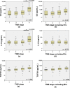
Cytokine concentrations in ascitic fluid according to TNM stage. (a) Peritoneal IL6 levels were higher in stage IV patients (p < 0.001 for stage III vs IV). (b) However, the peritoneal IL6 level of stage IV patients was not different if M1c patients were excluded (p = 0.170 for stage III vs IV). (c) The peritoneal IL10 level was higher in stage IV patients (p < 0.001 for stage III vs IV). (d) However, the peritoneal IL10 level of stage IV patients was not different if M1c patients were excluded (p = 0.860 for stage III vs IV). (e) Peritoneal transforming growth factor beta 1 (TGFB1) levels according to TNM stage (p = 0.130 for stage III vs IV). (f) TGFB1 levels according to TNM stage after excluding M1c patients (p = 0.532 for stage III vs IV). Ordinates are plotted on a log scale.
Figure 4
Figure 4
Ascitic IL10 level as a prognostic marker for peritoneal recurrence in patients with stage pT4 CRC. (a) Receiver operating characteristic curve for IL10 shows an AUC of 0.733 (p = 0.004). The relevant cut-off value is 14.0 pg/mL. (b) Peritoneal recurrence-free survival of patients with low (≤ 14.0 pg/mL) and high (> 14.0 pg/mL) peritoneal IL10 levels was significantly different (p < 0.001; log-rank test). AUC area under the curve, PR peritoneal recurrence.
Figure 5
Figure 5
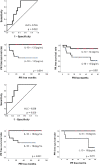
Classification performance of the IL10 cut-off model. (a) ROC curve from cohort I (n = 32) showed an AUC of 0.736 (p = 0.022). The relevant cut-off value was 13.5 pg/mL. (b) The difference in PR in cohort I was significant (p = 0.004; no PR of 13 for ≤ 13.5 pg/mL and 9 PR of 19 for > 13.5 pg/mL in cohort I). (c) The difference in PR in cohort II (n = 29) was also significant (p = 0.031; two PRs of 15 for ≤ 13.5 pg/mL and 7 PRs of 14 for > 13.5 pg/mL in cohort II). (d) The ROC curve from cohort I without SR (n = 21) showed an AUC of 0.838 (p = 0.026). The relevant cut-off value was 18.6 pg/mL. (e) The difference in PR in cohort I without SR was significant (p = 0.021; no PR of 10 for ≤ 18.6 pg/mL and 5 PR of 11 for > 18.6 pg/mL in cohort I). (f) The difference in PR in cohort II (n = 21) without SR was also significant (p = 0.011; no PR of 13 for ≤ 18.6 pg/mL and 3 PR of 8 for > 18.6 pg/mL in cohort II). ROC receiver operating characteristic, AUC area under the curve, PR peritoneal recurrence, SR systemic recurrence).

References

    1. Yamaguchim A, et al. Intraperitoneal hyperthermic treatment for peritoneal dissemination of colorectal cancers. Dis. Colon Rectum. 1992;35:964–968. doi: 10.1007/BF02253499. - DOI - PubMed
    1. Honore C, Goere D, Souadka A, Dumont F, Eliasm D. Definition of patients presenting a high risk of developing peritoneal carcinomatosis after curative surgery for colorectal cancer: A systematic review. Ann. Surg. Oncol. 2013;20:183–192. doi: 10.1245/s10434-012-2473-5. - DOI - PubMed
    1. Brodsky JT, Cohen AM. Peritoneal seeding following potentially curative resection of colonic carcinoma: Implications for adjuvant therapy. Dis. Colon Rectum. 1991;34:723–727. doi: 10.1007/BF02050360. - DOI - PubMed
    1. Mirnezami R, et al. Cytoreductive surgery in combination with hyperthermic intraperitoneal chemotherapy improves survival in patients with colorectal peritoneal metastases compared with systemic chemotherapy alone. Br. J. Cancer. 2014;111:1500–1508. doi: 10.1038/bjc.2014.419. - DOI - PMC - PubMed
    1. Bae SJ, et al. Role of peritoneal lavage cytology and prediction of prognosis and peritoneal recurrence after curative surgery for colorectal cancer. Ann. Coloproctol. 2014;30:266–273. doi: 10.3393/ac.2014.30.6.266. - DOI - PMC - PubMed

Publication types

MeSH terms