Review
. 2021 Jun;17(6):333-348.
doi: 10.1038/s41584-021-00605-2. Epub 2021 Apr 28.

Epithelial-immune cell interplay in primary Sjögren syndrome salivary gland pathogenesis

Affiliations
Review

Epithelial-immune cell interplay in primary Sjögren syndrome salivary gland pathogenesis

Gwenny M Verstappen et al. Nat Rev Rheumatol. 2021 Jun.

Abstract

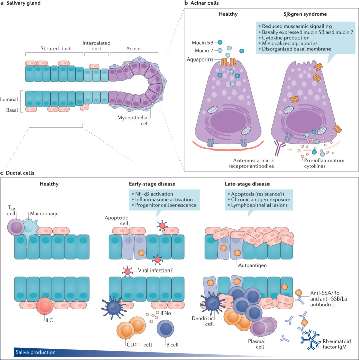
In primary Sjögren syndrome (pSS), the function of the salivary glands is often considerably reduced. Multiple innate immune pathways are likely dysregulated in the salivary gland epithelium in pSS, including the nuclear factor-κB pathway, the inflammasome and interferon signalling. The ductal cells of the salivary gland in pSS are characteristically surrounded by a CD4+ T cell-rich and B cell-rich infiltrate, implying a degree of communication between epithelial cells and immune cells. B cell infiltrates within the ducts can initiate the development of lymphoepithelial lesions, including basal ductal cell hyperplasia. Vice versa, the epithelium provides chronic activation signals to the glandular B cell fraction. This continuous stimulation might ultimately drive the development of mucosa-associated lymphoid tissue lymphoma. This Review discusses changes in the cells of the salivary gland epithelium in pSS (including acinar, ductal and progenitor cells), and the proposed interplay of these cells with environmental stimuli and the immune system. Current therapeutic options are insufficient to address both lymphocytic infiltration and salivary gland dysfunction. Successful rescue of salivary gland function in pSS will probably demand a multimodal therapeutic approach and an appreciation of the complicity of the salivary gland epithelium in the development of pSS.


Conflict of interest statement

The authors declare no competing interests.

Figures

Fig. 1
Fig. 1. The salivary gland epithelium in health and in pSS.
a | The salivary gland epithelium is made up of several cell types, including acinar cells (that cluster to form the acinus), ductal cells (consisting of basal and luminal cell types) that form the striated and intercalated ducts, myoepithelial cells and progenitor cells. Saliva is produced by acinar cells and secreted upon myoepithelial cell contraction. Saliva is then channelled and modified through the intercalated and striated ducts and flows via larger excretory ducts into the mouth. b | Acinar cells contain multiple defects in primary Sjögren syndrome (pSS). c | A number of pathogenic events could take place in the ductal epithelium during pSS development. In a healthy situation, several immune cells (for example, macrophages, innate lymphoid cells (ILCs) and tissue resident memory (TRM) cells) are present for immune surveillance to enable a fast response to injury or infection. In pSS, an unknown trigger (for example, viral infection or tissue damage) might cause activation of innate immune pathways, epithelial cell apoptosis and senescence, which is exacerbated in more severe stages of disease. Late-stage disease is characterized by chronic antigen exposure, local production of autoantibodies, accumulation of CD4+ T cells and B cells, complete loss of progenitor cells and the formation of lymphoepithelial lesions. These lesions are characterized by basal cell hyperplasia and the presence of intraepithelial lymphocytes.
Fig. 2
Fig. 2. Lymphoepithelial lesions in the salivary glands of a patient with primary Sjögren syndrome.
Consecutive sections of parotid gland tissue from a patient with primary Sjögren syndrome are shown. Lymphoepithelial lesions are indicated by an asterisk on the haematoxylin and eosin (H&E)-stained section. Consecutive sections were stained with antibodies to reveal either epithelial cells (using antibodies against high molecular weight cytokeratin (hmwCK)), B cells (using antibodies against CD20) or T cells (using antibodies against CD3). B cells dominate the lymphoepithelial lesions, whereas T cells are rarely detected within these structures. Image courtesy of M.S. van Ginkel and B. van der Vegt.
Fig. 3
Fig. 3. Interactions between FcRL4+ B cells and the ductal epithelium in primary Sjögren syndrome.
a | Activated epithelial cells secrete CXCL10 and attract Fc-receptor like 4-positive (FcRL4+) B cells. These cells are further activated by B cell-activating factor (BAFF) and a proliferation-inducing ligand (APRIL), secreted by epithelial cells, and other stimuli from the pro-inflammatory environment. Upon activation, FcRL4+ B cells express CD11c, which can form an integrin together with the β2 integrin. This integrin can bind to intercellular adhesion molecule 1 (ICAM1) on epithelial cells and sustain their interaction. The production of IL-6 and possibly also other pro-inflammatory cytokines by FcRL4+ B cells might result in hyperplasia of the epithelium.

References

    1. Sjögren H. Zur Kenntnis der Keratoconjunctivitis sicca (Keratitis filiformis bei Hypofunktion der Tränendrüsen) Acta Ophthalmol. 1933;11(Suppl 2):1–51.
    1. Brito-Zerón P, et al. Sjögren syndrome. Nat. Rev. Dis. Primers. 2016;2:16047. - PubMed
    1. Shiboski CH, et al. 2016 American College of Rheumatology/European League Against Rheumatism classification criteria for primary Sjögren’s syndrome: a consensus and data-driven methodology involving three international patient cohorts. Arthritis Rheumatol. 2017;69:35–45. - PMC - PubMed
    1. Greenspan JS, Daniels TE, Talal N, Sylvester RA. The histopathology of Sjögren’s syndrome in labial salivary gland biopsies. Oral Surg. Oral Med. Oral Pathol. 1974;37:217–229. - PubMed
    1. Christodoulou MI, Kapsogeorgou EK, Moutsopoulos HM. Characteristics of the minor salivary gland infiltrates in Sjogren’s syndrome. J. Autoimmun. 2010;34:400–407. - PubMed

Publication types

MeSH terms