. 2021 Jan-Feb;66(1):24-33.
doi: 10.4103/ijd.IJD_602_20.

Post Kala-Azar Dermal Leishmaniasis: Clinical Features and Differential Diagnosis

Affiliations

Post Kala-Azar Dermal Leishmaniasis: Clinical Features and Differential Diagnosis

Piyush Kumar et al. Indian J Dermatol. 2021 Jan-Feb.

Abstract

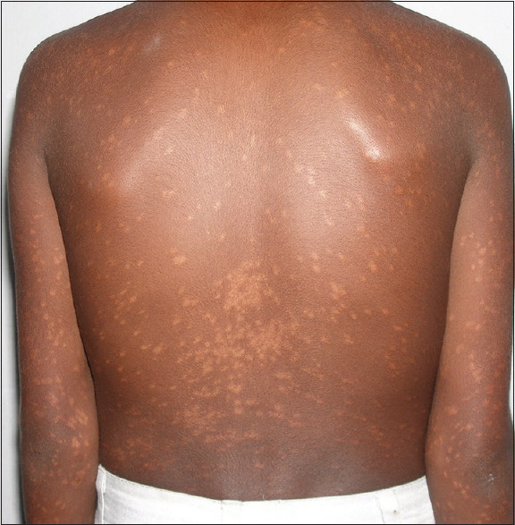
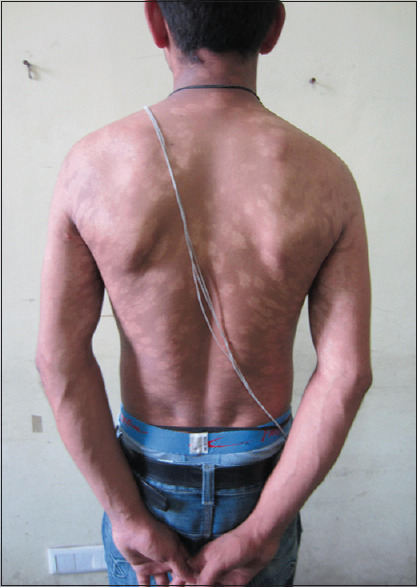
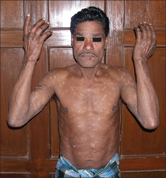
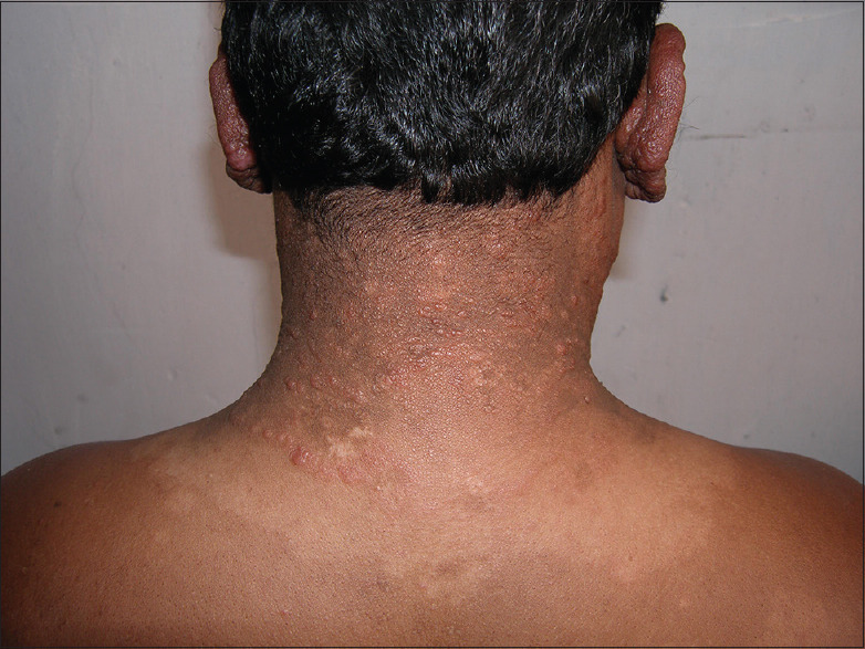
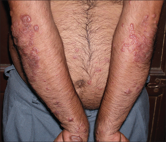
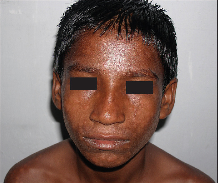
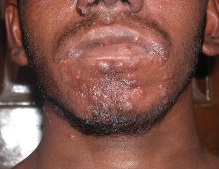
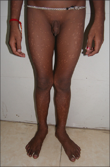
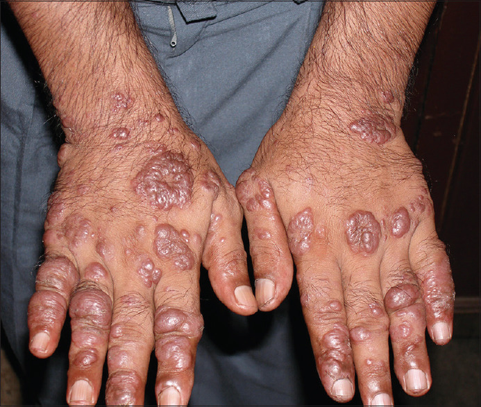
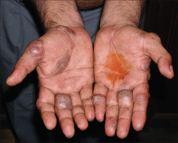
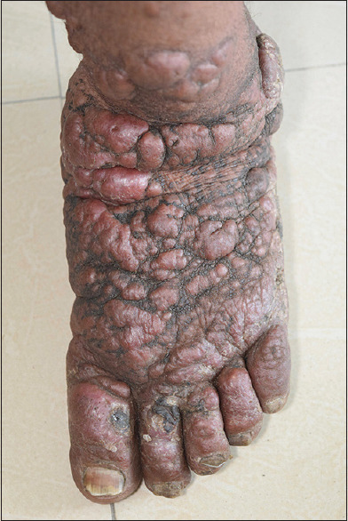
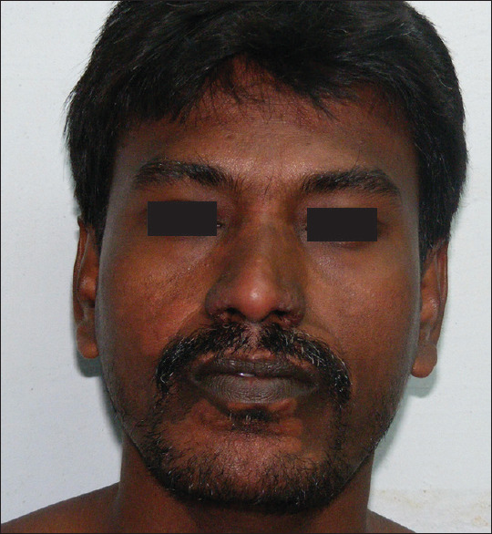
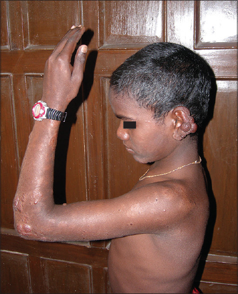
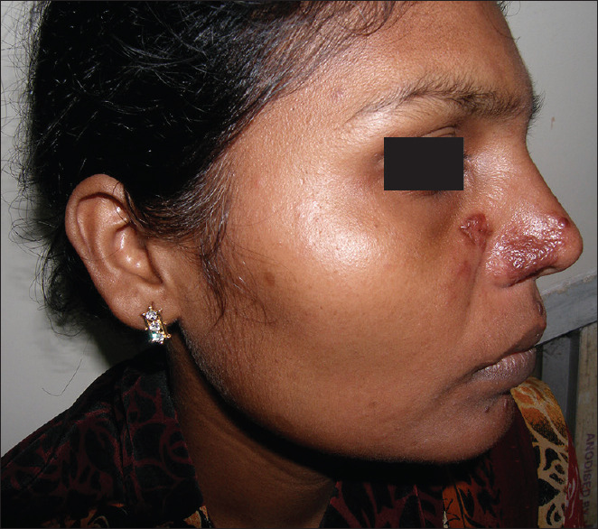
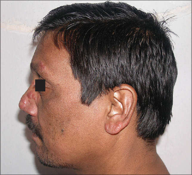
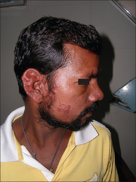
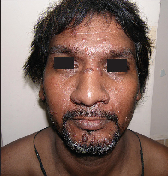
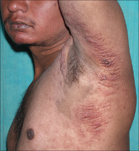
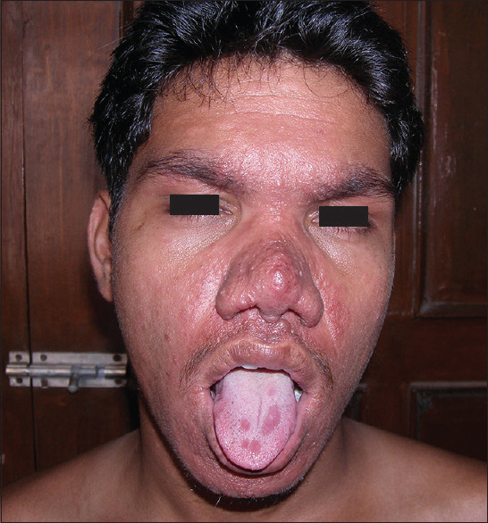
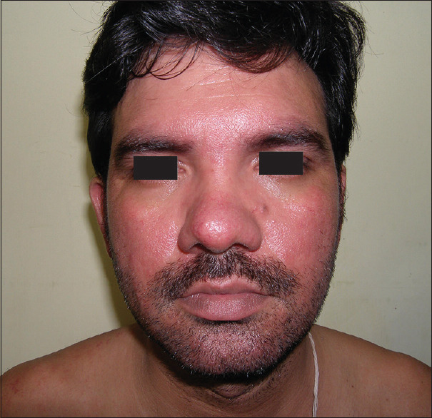
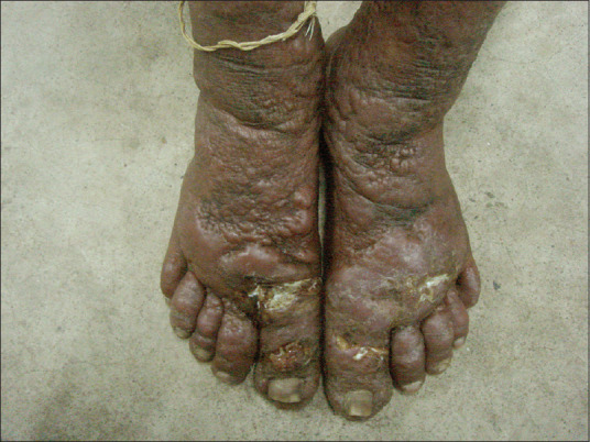
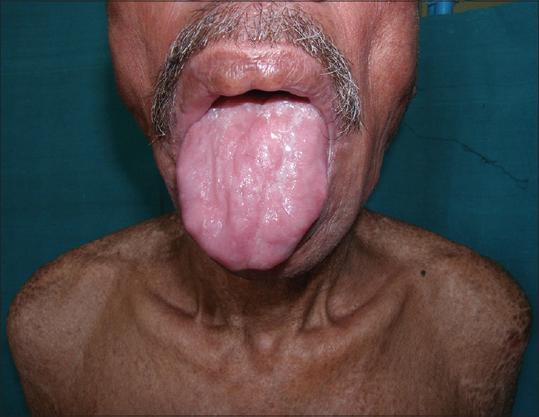
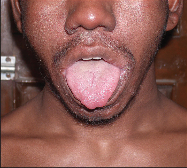
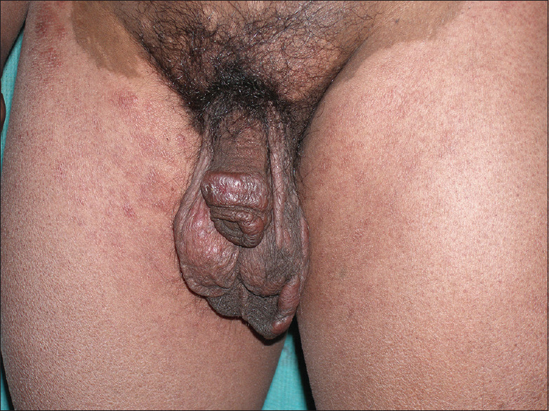
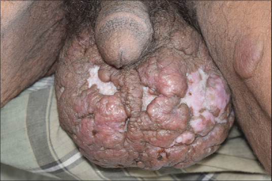
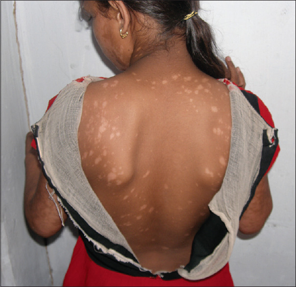
Post kala-azar dermal leishmaniasis (PKDL) is a mucocutaneous disease usually seen in apparently cured, inadequately treated or untreated cases of visceral leishmaniasis and is endemic to many parts of India, Nepal, Bangladesh, and eastern Africa (Sudan, Ethiopia, Kenya). The disease usually manifests as a variable combination of hypopigmented patches, erythematous succulent papulo-plaques, and nodular lesions on the face and upper body and sometimes extending on the extremities, genitalia, and tongue. Atypical morphology and presentations are not uncommon, especially in endemic areas, which include photosensitivity, verrucous, hypertrophic, xanthomatous, and ulcerative lesions. Recognition of spectrum of mucocutaneous changes helps physicians in early initiation of treatment and in reducing disease transmission in the community. The differential diagnosis depends on the pattern of manifestations, but lepromatous leprosy is the closest mimicker. Since PKDL does not cause significant morbidity, at least initially, but the affected patients continue to act as a reservoir of the disease, active case detection is required to identify cases early to control the disease transmission in the community.

Keywords: Active surveillance; India; Leishmania donovani; Sudan; para kala-azar dermal leishmaniasis; post kala-azar dermal leishmaniasis; visceral leishmaniasis.


Conflict of interest statement

There are no conflicts of interest.

Figures

Figure 1
Figure 1
Monomorphic macular lesions of PKDL on the trunk
Figure 2
Figure 2
Monomorphic macular lesions which have coalesced together
Figure 3
Figure 3
Polymorphic lesions with guttate hypopigmented macules on trunk, plaques on left forearm, and nodules on abdomen
Figure 4
Figure 4
Polymorphic lesions with macules on back, papules on nape of neck, and nodules on earlobe
Figure 5
Figure 5
Papular and nodular lesions developing de-novo near the elbow and wrist
Figure 6
Figure 6
Macules developing around “muzzle area of face” and then spreading to rest of face
Figure 7
Figure 7
Papules and nodules on the “muzzle-area of face”
Figure 8
Figure 8
Macules spreading on the lower extremities
Figure 9
Figure 9
Nodules spreading on the dorsa of hands
Figure 10
Figure 10
Nodules extending onto the palms
Figure 11
Figure 11
Nodules on the foot
Figure 12
Figure 12
Papule developing over a patch near the lower lip
Figure 13
Figure 13
Nodules developing over the patch near the elbow
Figure 14
Figure 14
Papules and succulent nodules over the nose
Figure 15
Figure 15
Papules and succulent nodules over the nose and ear
Figure 16
Figure 16
Succulent nodules over ears
Figure 17
Figure 17
Papules and succulent nodules over the entire face
Figure 18
Figure 18
Hypopigmented patches and papules occupying the anterior and posterior axillary folds but spare a small island of skin near the vault of axilla
Figure 19
Figure 19
Photosensitivity with nodules on nose and tongue
Figure 20
Figure 20
Photosensitivity was the presenting feature in the case who went on to develop hypopigmented patches later
Figure 21
Figure 21
Spontaneous ulceration developing on top of nodules in PKDL which became secondarily infected
Figure 22
Figure 22
Nodules on the tongue and upper lip
Figure 23
Figure 23
Nodules on the lips and tip of tongue
Figure 24
Figure 24
Nodules on the genitals with confluent hypopigmented patches on the thighs
Figure 25
Figure 25
Nodules on the genitals
Figure 26
Figure 26
Hypopigmented macules on the covered area of back in a young girl

References

    1. Brahmachari UN. A new form of cutaneous Leishmaniasis-Dermal Leishmanoid. Ind Med Gaz. 1922;57:125–7. - PMC - PubMed
    1. Das Gupta BM. A note on the parasite of “Dermal Leishmanoid”. Ind Med Gaz. 1927;62:11–2. - PMC - PubMed
    1. Gupta BMD. Further observations upon “Dermal Leishmanoid”. Ind Med Gaz. 1927;62:199. - PMC - PubMed
    1. Seshadrinathan N. Report of a second case of “Dermal Leishmanoid” from Madras. Ind Med Gaz. 1930;65:567–8. - PMC - PubMed
    1. Theodore JH. Note on a case of “Dermal Leishmanoid” from Madras. Ind Med Gaz. 1930;65:508. - PMC - PubMed

LinkOut - more resources