

Isolated Plantar Vein Thrombosis Resembling a Corn with a Bruise
- PMID: 33911541
- PMCID: PMC7992697
- DOI: 10.5021/ad.2019.31.1.66
Isolated Plantar Vein Thrombosis Resembling a Corn with a Bruise
Abstract
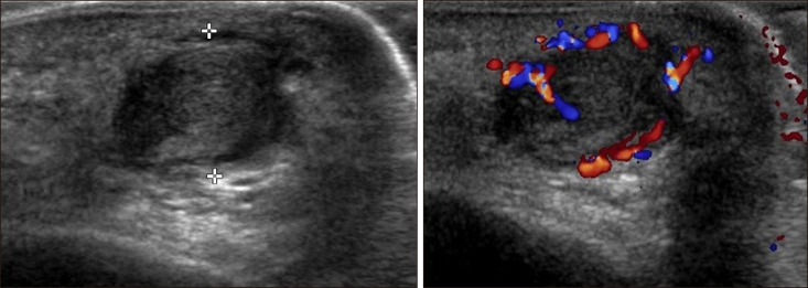
Plantar vein thrombosis, rarely-reported disease, is usually accompanied by pain and tenderness in the plantar region and should be differentiated from other dermatological conditions causing plantar pain, such as hemorrhagic corn/callus, plantar epidermal cyst, verruca, or plantar fibromatosis. A 52-year-old man presented with a violaceous tender subcutaneous nodule overlying a hyperkeratotic plaque on his sole. Initially, he thought it was a corn and applied keratolytic agents, which failed to work. Sonography revealed a well-demarcated mass with increased peripheral vascularity. His pain was relieved after a complete wide excision, which confirmed the mass to be plantar vein thrombosis after histopathological examination.
Keywords: Corn; Plantar vein thrombosis; Venous thrombosis.
Copyright © 2019 The Korean Dermatological Association and The Korean Society for Investigative Dermatology.
Conflict of interest statement
CONFLICTS OF INTEREST: The authors have nothing to disclose.
Figures


Similar articles
-
Clinics in diagnostic imaging (195). Plantar fibromatosis.Singapore Med J. 2019 May;60(5):230-235. doi: 10.11622/smedj.2019043. Singapore Med J. 2019. PMID: 31187147 Free PMC article.
-
Antiphospholipid Syndrome with Antiβ2glicoprotein-1 Antibodies as the Cause of Recurrent Tibial Vein Thrombosis in SAPHO syndrome.Acta Dermatovenerol Croat. 2016 Dec;24(4):305-306. Acta Dermatovenerol Croat. 2016. PMID: 28128085
-
Clinical characteristics and course of plantar vein thrombosis: a series of 22 cases.Phlebology. 2015 Dec;30(10):714-8. doi: 10.1177/0268355514555385. Epub 2014 Oct 17. Phlebology. 2015. PMID: 25326214
-
Spontaneous plantar vein thrombosis: state of the art.Phlebology. 2013 Dec;28(8):432-7. doi: 10.1177/0268355513477087. Epub 2013 May 6. Phlebology. 2013. PMID: 23520215 Review.
-
Mesenteric cysts and mesenteric venous thrombosis leading to intestinal necrosis in pregnancy managed with laparotomy: a case report and review of the literature.J Med Case Rep. 2017 Jul 7;11(1):184. doi: 10.1186/s13256-017-1320-5. J Med Case Rep. 2017. PMID: 28683785 Free PMC article. Review.
References
-
- Wallace GF. Dermatologic causes of heel pain. Clin Podiatr Med Surg. 2010;27:407–416. - PubMed
-
- Bernathova M, Bein E, Bendix N, Bodner G. Sonographic diagnosis of plantar vein thrombosis: report of 3 cases. J Ultrasound Med. 2005;24:101–103. - PubMed
-
- Siegal DS, Wu JS, Brennan DD, Challies T, Hochman MG. Plantar vein thrombosis: a rare cause of plantar foot pain. Skeletal Radiol. 2008;37:267–269. - PubMed
-
- Long A, Bura-Riviere A, Sapoval M. [Plantar venous thrombosis and anticardiolipin antibody syndrome. Case report] J Mal Vasc. 2004;29:39–40. French. - PubMed
-
- Cavezzi A. Isolated thrombosis of plantar veins. Case report. Minerva Cardioangiol. 1999;47:309–313. - PubMed
Publication types
LinkOut - more resources
Full Text Sources
