

Syringocystadenocarcinoma Papilliferum: A Case Report and Review of the Literature
- PMID: 33911649
- PMCID: PMC7992554
- DOI: 10.5021/ad.2019.31.5.559
Syringocystadenocarcinoma Papilliferum: A Case Report and Review of the Literature
Abstract
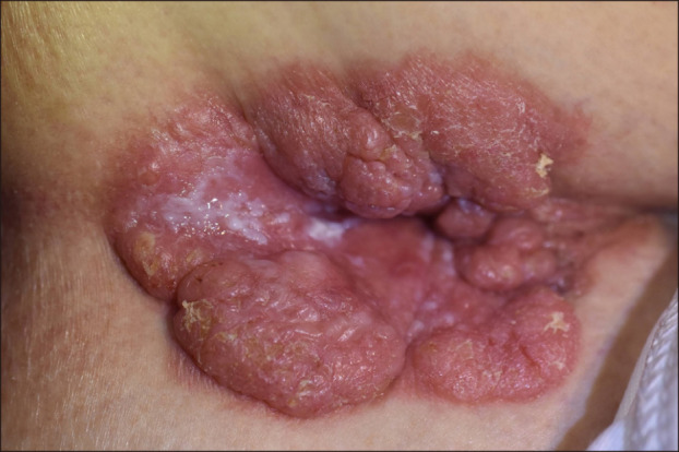
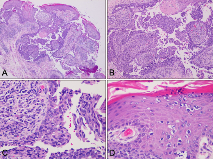
Syringocystadenocarcinoma papilliferum (SCACP) is a rare malignant adnexal neoplasm, which is considered as a malignant counterpart of syringocystadenoma papilliferum (SCAP). Clinically, SCACP appears as a nodule, inflammatory plaque, or tumor. The lesion is usually covered with crusts, which are formed by secretion of the apocrine epithelial cells. Histologically, SCACP resembles SCAP, with cystic papillomatous invaginations connected to the skin surface by funnel-shaped structures lined by infundibular epithelium. The stroma of the tumor consists of a dense inflammatory infiltrate of plasma cells and lymphocytes. SCACP differs from SCAP in terms of the architectural and cytological features of the tumor cells, and is characterized by higher nuclear cytoplasmic ratio, nuclear irregularity, coarse chromatin, and increased mitotic activity. However, the immunohistochemical findings of SCACP vary. Since only 49 cases of SCACP have been reported in the English literature, the clinical and histologic characteristics of SCACP have not been fully established. Further studies on the diagnostic criteria for SCACP are warranted. Here, we report a rare case of SCACP and present a review of other relevant literature.
Keywords: Sweat gland neoplasm; Syringocystadenocarcinoma papilliferum; Syringocystadenoma papilliferum.
Copyright © 2019 The Korean Dermatological Association and The Korean Society for Investigative Dermatology.
Conflict of interest statement
CONFLICTS OF INTEREST: The authors have nothing to disclose.
Figures
References
-
- Numata M, Hosoe S, Itoh N, Munakata Y, Hayashi S, Maruyama Y. Syringadenocarcinoma papilliferum. J Cutan Pathol. 1985;12:3–7. - PubMed
-
- Chen J, Beg M, Chen S. Syringocystadenocarcinoma papilliferum in situ, a variant of cutaneous adenocarcinoma in situ: a case report with literature review. Am J Dermatopathol. 2016;38:762–765. - PubMed
-
- Parekh V, Guerrero CE, Knapp CF, Elmets CA, McKay KM. A histological snapshot of hypothetical multistep progression from nevus sebaceus to invasive syringocystadenocarcinoma papilliferum. Am J Dermatopathol. 2016;38:56–62. - PubMed
-
- Leeborg N, Thompson M, Rossmiller S, Gross N, White C, Gatter K. Diagnostic pitfalls in syringocystadenocarcinoma papilliferum: case report and review of the literature. Arch Pathol Lab Med. 2010;134:1205–1209. - PubMed
Publication types
LinkOut - more resources
Full Text Sources
