. 2021 Apr 12:12:638469.
doi: 10.3389/fimmu.2021.638469. eCollection 2021.

Subversion of Host Innate Immunity by Rickettsia australis via a Modified Autophagic Response in Macrophages

Affiliations

Subversion of Host Innate Immunity by Rickettsia australis via a Modified Autophagic Response in Macrophages

Jeremy Bechelli et al. Front Immunol. .

Abstract

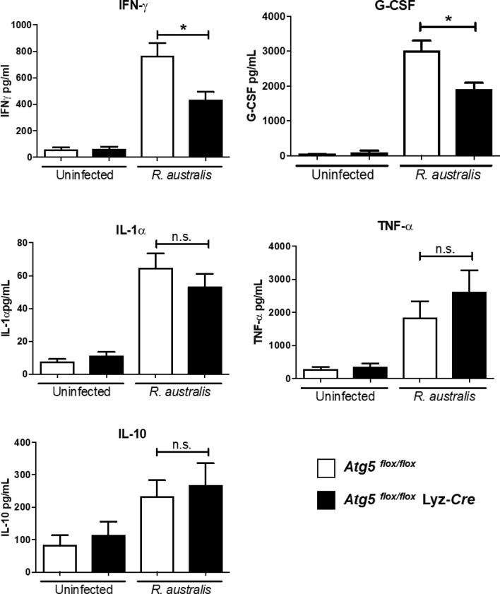
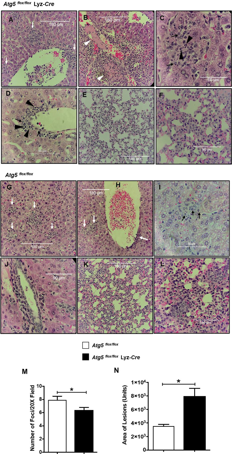
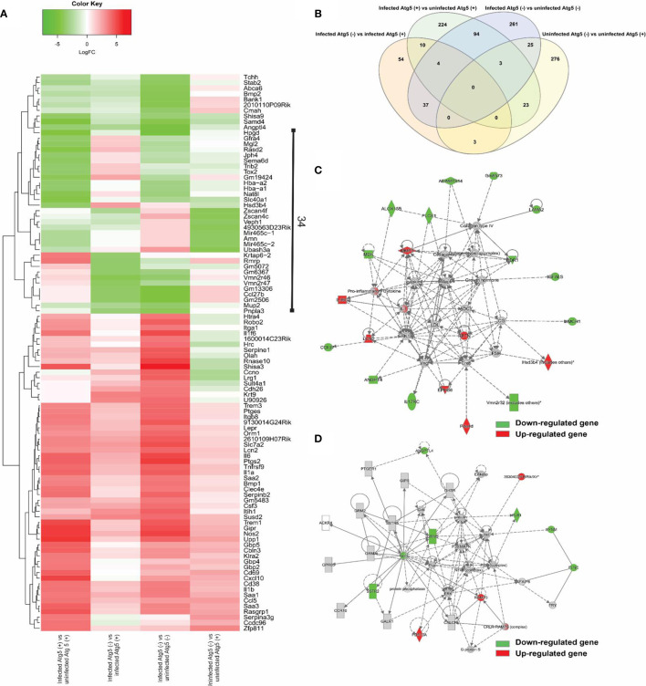
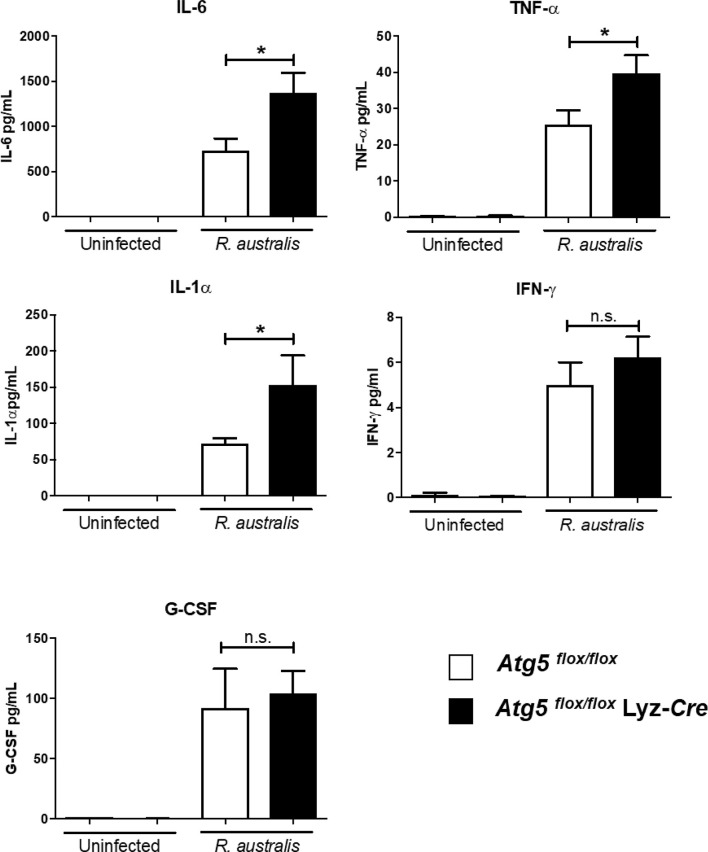
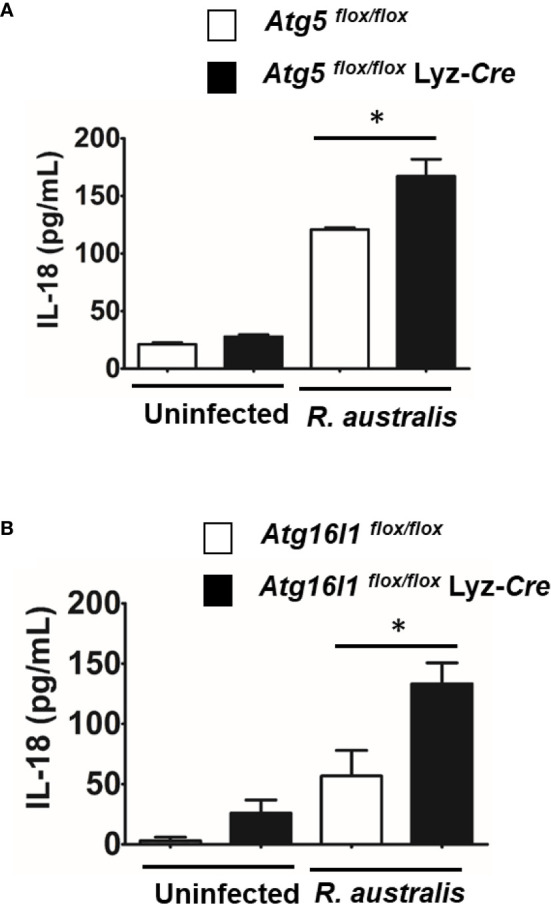
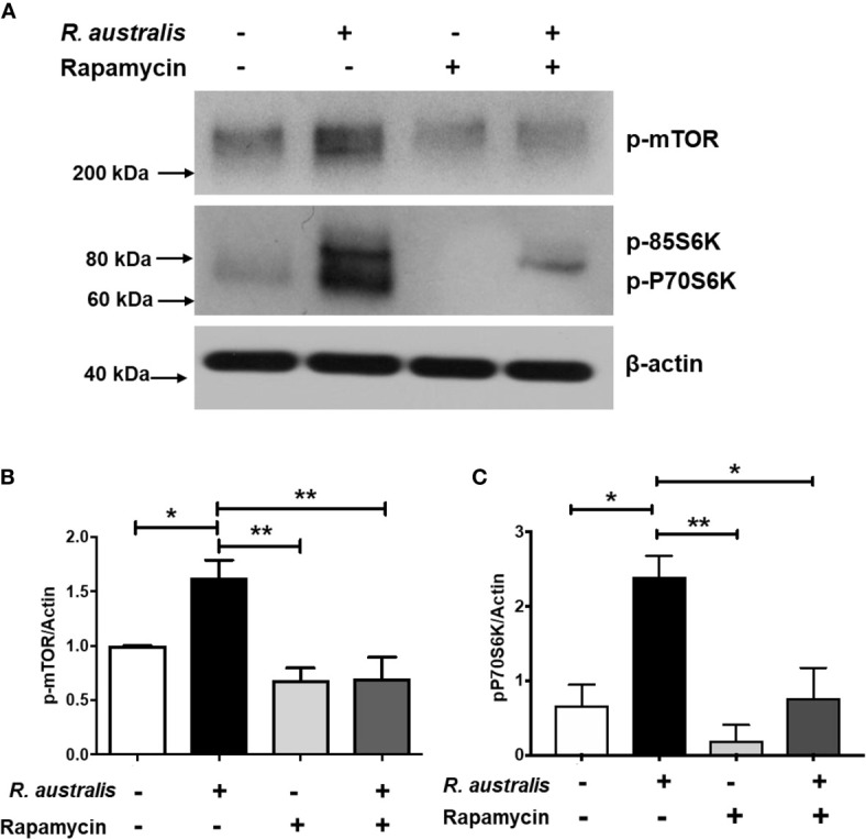
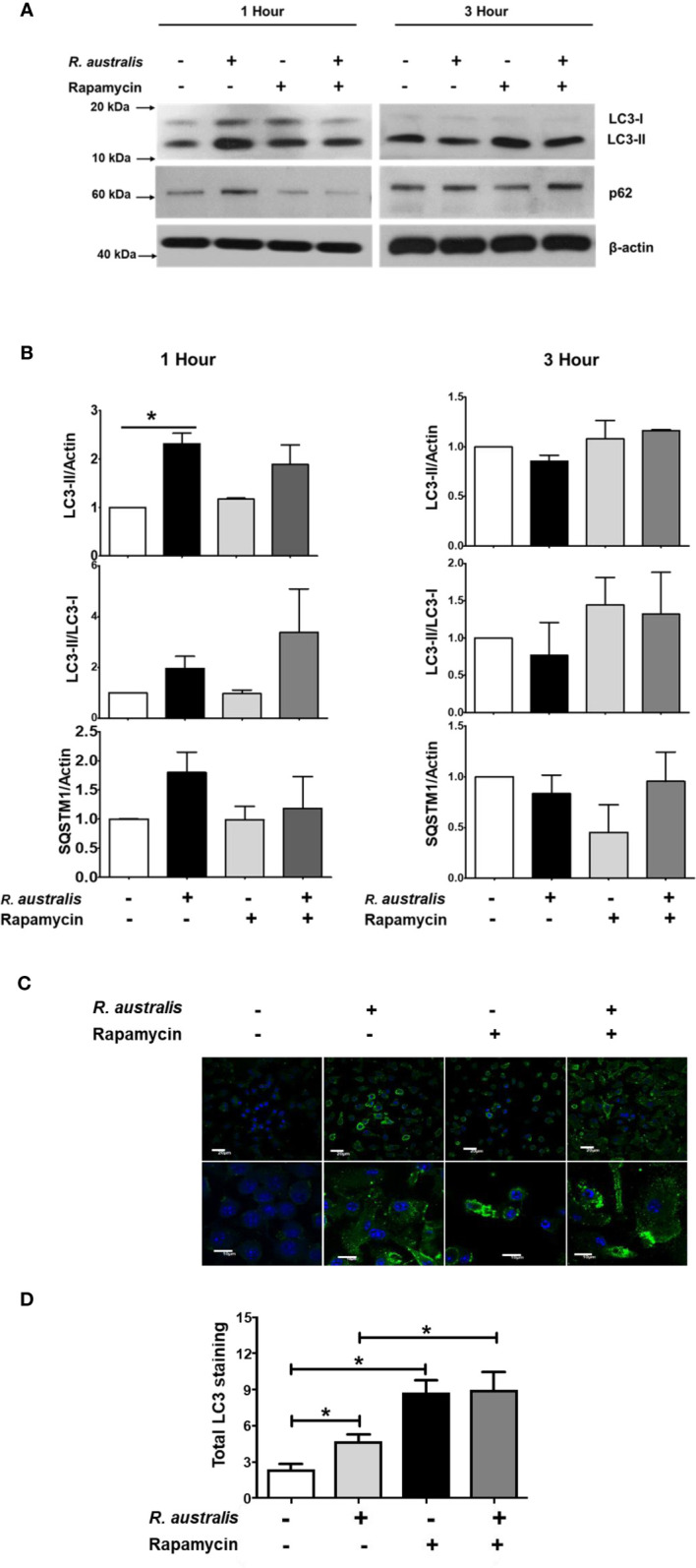
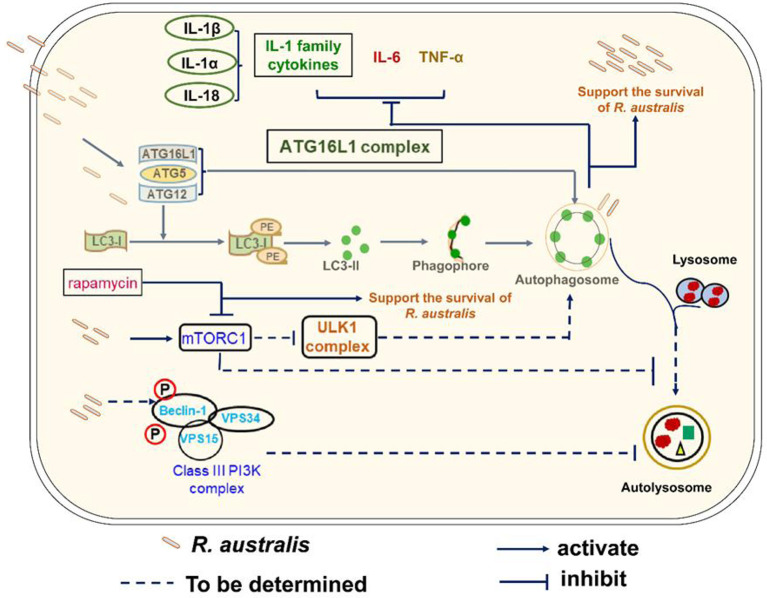
We recently reported that the in vitro and in vivo survivals of Rickettsia australis are Atg5-dependent, in association with an inhibited level of anti-rickettsial cytokine, IL-1β. In the present study, we sought to investigate how R. australis interacts with host innate immunity via an Atg5-dependent autophagic response. We found that the serum levels of IFN-γ and G-CSF in R. australis-infected Atg5flox/flox Lyz-Cre mice were significantly less compared to Atg5flox/flox mice, accompanied by significantly lower rickettsial loads in tissues with inflammatory cellular infiltrations including neutrophils. R. australis infection differentially regulated a significant number of genes in bone marrow-derived macrophages (BMMs) in an Atg5-depdent fashion as determined by RNA sequencing and Ingenuity Pathway Analysis, including genes in the molecular networks of IL-1 family cytokines and PI3K-Akt-mTOR. The secretion levels of inflammatory cytokines, such as IL-1α, IL-18, TNF-α, and IL-6, by R. australis-infected Atg5flox/flox Lyz-Cre BMMs were significantly greater compared to infected Atg5flox/flox BMMs. Interestingly, R. australis significantly increased the levels of phosphorylated mTOR and P70S6K at a time when the autophagic response is induced. Rapamycin treatment nearly abolished the phosphorylated mTOR and P70S6K but did not promote significant autophagic flux during R. australis infection. These results highlight that R. australis modulates an Atg5-dependent autophagic response, which is not sensitive to regulation by mTORC1 signaling in macrophages. Overall, we demonstrate that R. australis counteracts host innate immunity including IL-1β-dependent inflammatory response to support the bacterial survival via an mTORC1-resistant autophagic response in macrophages.

Keywords: Rickettsia; autophagy; innate immunity; mTOR signaling; macrophages.


Conflict of interest statement

The authors declare that the research was conducted in the absence of any commercial or financial relationships that could be construed as a potential conflict of interest.

Figures

Figure 1
Figure 1
(previous Figure 6). R. australis subverts host innate immunity against rickettsioses via Atg5-dependent autophagic response. Atg5 flox/flox Lyz-Cre and Atg5 flox/flox mice were infected with R. australis i.v. at a dose of 3 × 105 PFU per mouse. On day 4 p.i., mice were euthanized, and serum was collected. Systemic production levels of cytokines/chemokines including IFN-γ, G-CSF, TNF-α, IL-1α, and IL-10 in mouse serum were determined by Bioplex assay. Results are means ± SE of data from three independent experiments containing 4-6 mice per group. *p<0.05; n.s., not statistically significant.
Figure 2
Figure 2
(previous Figure 7). Inflammatory cellular accumulation upon infection with R. australis in tissues of Atg5 flox/flox mice and Atg5 flox/flox Lyz-Cre mice. Mice were infected i.v. with R. australis (3 × 10⁵ PFU per mouse). On day 4 p.i., mice were sacrificed, and tissues were isolated and analyzed. Histological analysis of livers and lungs from infected Atg5 flox/flox Lyz-Cre mice (A–F) and Atg5 flox/flox mice (G–L). Foci of inflammatory infiltration are indicated by white arrows. Thrombus or necrotic cells related to thrombosis is shown as white arrowheads (B). Polymorphonuclear neutrophils (PMNs) (black arrowheads) and macrophages (black arrows) are shown in livers. Furthermore, the size (M) and frequency (N) of inflammatory lesions in livers were analyzed using ImageJ (magnification, ×20). Images were taken using an Olympus BX41 photomicroscope (Olympus America, Inc., Center Valley, PA) or using a Revolution Microscope and an iPad Pro® tablet (Echo Laboratory, San Diego, CA). *p<0.05.
Figure 3
Figure 3
(previous Figure 3). Comparative transcriptional analysis of R. australis-infected Atg5 flox/flox Lyz-Cre and Atg5 flox/flox BMMs. BMMs were isolated from Atg5 flox/flox Lyz-Cre and Atg5 flox/flox mice, and then infected with R. australis at an MOI of 5. At 24 h p.i., cells were collected and total RNA was extracted. RNA-seq analysis was performed as described in Materials and Methods. Atg5 (+), Atg5 flox/flox ; Atg5 (-), Atg5 flox/flox Lyz-Cre. (A), Heatmap and hierarchical clustering of the top 100 genes differentially regulated by Atg5 during R. australis infection in mouse macrophages. The expression levels of genes are indicated by the color bar above the heatmap. Red color indicates the increased expression whereas green color indicates the decreased expression in four comparisons. (B), Venn diagram showing overlap of significantly modulated genes for each of the four comparisons. (C, D), IPA molecular networks analysis of differentially expressed genes in IL-1 family cytokines signaling and PI3K-Akt-mTOR signaling in R. australis-infected Atg5 flox/flox Lyz-Cre BMMs vs R. australis-infected Atg5 flox/flox BMMs. Red, up-regulated; green, down-regulated.
Figure 4
Figure 4
(previous Figure 4). R. australis counteracts the production of pro-inflammatory cytokines by infected macrophages via Atg5-dependent autophagy. BMMs were isolated from Atg5 flox/flox Lyz-Cre and Atg5 flox/flox mice, and they were infected with R. australis at an MOI of 5. At 24 h p.i., supernatant was harvested. Production levels of cytokines including IL-6, TNF-α, IL-1α, IFN-γ and G-CSF in the supernatant were assessed by Bioplex assay. Results are means ± SE of data from three independent experiments. *p<0.05; n.s., not statistically significant.
Figure 5
Figure 5
(previous Figure 5). R. australis suppressed the production of IL-18 by infected macrophages via Atg5-dependent autophagy. BMMs were isolated from Atg5 flox/flox Lyz-Cre and Atg5 flox/flox mice, and Atg16l1 flox/flox Lyz-Cre and Atg16l1 flox/flox mice. Macrophages were infected with R. australis at an MOI of 5. At 24 h p.i., supernatant was harvested. Concentrations of IL-18 in the supernatant of infected Atg5 flox/flox Lyz-Cre BMMs and Atg5 flox/flox BMMs (A), and infected Atg16l1 flox/flox Lyz-Cre BMMs and Atg16l1 flox/flox BMMs (B) were assessed by ELISA. Results are means ± SE of data from two independent experiments. *p < 0.05.
Figure 6
Figure 6
(previous Figure 1). R. australis activates mTORC1 in mouse macrophages. BMMs of WT B6 mice were isolated and infected with R. australis at an MOI of 5. To inhibit mTORC1 signaling, cells were treated with 50 ng/mL of rapamycin for 4 hours prior to infection with R. australis. At 1 h p.i., cells were collected, and cell lysates were immunoblotted with antibodies directed against phosphorylated mTOR, phosphorylated p70S6K, and β-actin (A). The activation of phosphorylated mTOR (B) and phosphorylated p70S6K (C) was analyzed by densitometry using β-actin as a normalization control with three independent replicates. *p < 0.05; **p < 0.01.
Figure 7
Figure 7
(previous Figure 2). Interactions of mTORC1 and autophagy with R. australis in macrophages. BMMs of WT B6 mice were isolated and infected with R. australis at an MOI of 5. To inhibit mTORC1 signaling, cells were treated with 50 ng/mL of rapamycin for 4 hours prior to infection with R. australis. Cells were collected at 1 h and 3 h p.i., and cell lysates were immunoblotted with antibodies directed against LC3-II, p62 and β-actin (A). (B), The ratios of LC3-II/Actin, LC3-II/LC3-I, and SQSTM1/Actin in uninfected and infected BMMs with or without rapamycin treatment at 1 h and 3 h p.i. were analyzed by densitometry. (C), Representative confocal immunofluorescence microscopic images of uninfected and infected BMMs with or without rapamycin treatment at 1 h p.i. at a magnification of 20x. Green, LC3 puncta; blue, nuclei (DAPI). Bar = 20 µm in the upper and 10 µm in the bottom row, respectively. (D), Total LC3 staining was quantified using Image J software. Microscopy data represent two to three independent experiments. Data shown are mean ± SE. Group comparison was not labeled if not statistically significant. *p < 0.05.
Figure 8
Figure 8
Schematic diagram of interactions of Atg5-dependent autophagy and mTORC1 with R. australis in macrophages. This schematic diagram depicts the molecular mechanisms involved in the modulation of autophagy and activation of mTORC1 by R. australis in order to benefit bacterial infection. R. australis activates the ATG5-ATG12-ATG16L1 complex leading to accumulation of LC3 (+) autophagosomes. Atg5 (+) LC3 (+) autophagosomes induced by R. australis in macrophages inhibit production levels of IL-1 family cytokines including IL-1β, IL-1α and IL-18 as well as other inflammatory cytokines such as IL-6 and TNF-α, the effects of which favor rickettsial infection. R. australis activates mTORC1 signaling at 1 h p.i. in macrophages when the Atg5-dependent autophagic response is induced. Thus, R. australis-induced autophagic response is resistant to regulation by mTORC1 signaling. It would be interesting to explore whether R. australis-induced autophagosomes can fuse with lysosome and mature into autolysosomes, and how ULK and class III PI3K complex are involved in this process in the future. Solid lines represent pathways demonstrated in our current and previous studies, while dashed lines refer to the mechanisms hypothesized but not tested.

References

    1. Straily A, Drexler N, Cruz-Loustaunau D, Paddock CD, Alvarez-Hernandez G. Notes from the Field: Community-based prevention of Rocky Mountain spotted fever - Sonora, Mexico, 2016. MMWR Morb Mortal Wkly Rep (2016) 65(46):1302–3. 10.15585/mmwr.mm6546a6 - DOI - PubMed
    1. Sahni SK, Rydkina E. Progress in the functional analysis of rickettsial genes through directed mutagenesis of Rickettsia prowazekii phospholipase D. Future Microbiol (2009) 4(10):1249–53. 10.2217/fmb.09.99 - DOI - PubMed
    1. Rumfield C, Hyseni I, McBride JW, Walker DH, Fang R. Activation of ASC inflammasome driven by Toll-like receptor 4 contributes to host immunity against rickettsial Infection. Infect Immun (2020) 88(4):e00886–19. 10.1128/IAI.00886-19 - DOI - PMC - PubMed
    1. Bechelli J, Vergara L, Smalley C, Buzhdygan TP, Bender S, Zhang W, et al. . Atg5 supports Rickettsia australis infection in macrophages in vitro and in vivo . Infect Immun (2018) 87(1):e00651–18. 10.1128/IAI.00651-18 - DOI - PMC - PubMed
    1. Smalley C, Bechelli J, Rockx-Brouwer D, Saito T, Azar SR, Ismail N, et al. . Rickettsia australis activates inflammasome in human and murine macrophages. PloS One (2016) 11(6):e0157231. 10.1371/journal.pone.0157231 - DOI - PMC - PubMed

Publication types

Supplementary concepts