. 2021 Apr 12:12:640842.
doi: 10.3389/fimmu.2021.640842. eCollection 2021.

Vasculitis and Neutrophil Extracellular Traps in Lungs of Golden Syrian Hamsters With SARS-CoV-2

Affiliations

Vasculitis and Neutrophil Extracellular Traps in Lungs of Golden Syrian Hamsters With SARS-CoV-2

Kathrin Becker et al. Front Immunol. .

Abstract

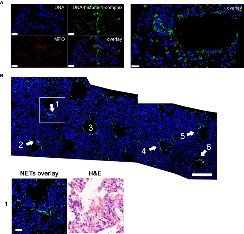
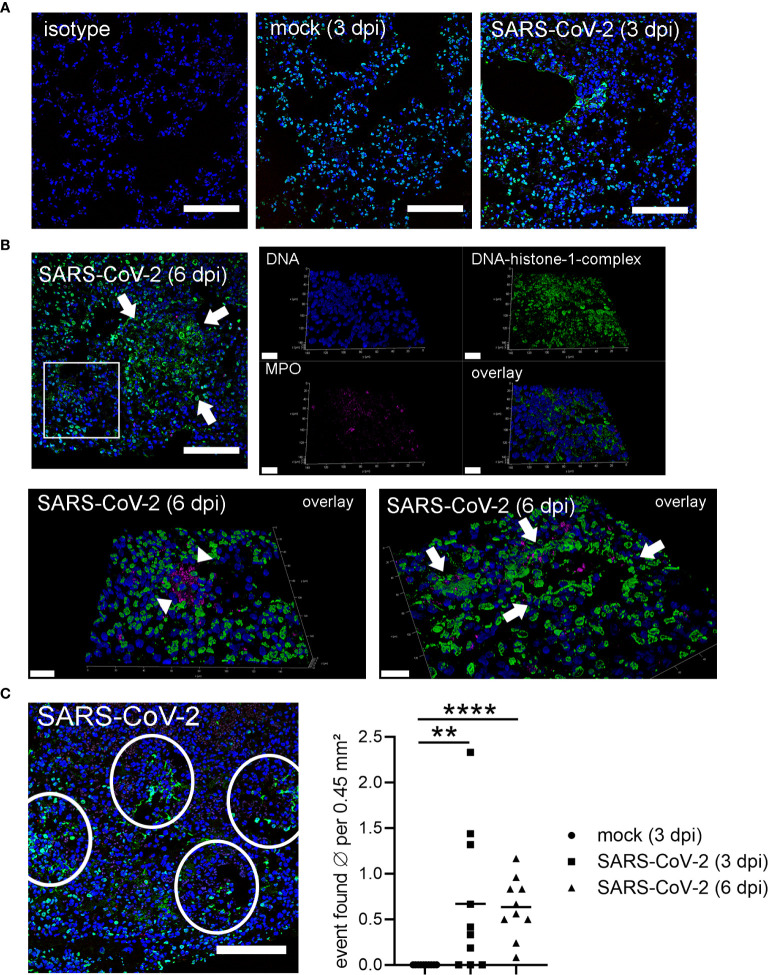
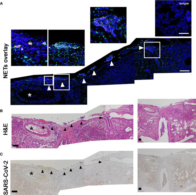
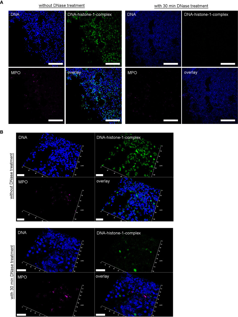
Neutrophil extracellular traps (NETs) have been identified as one pathogenetic trigger in severe COVID-19 cases and therefore well-described animal models to understand the influence of NETs in COVID-19 pathogenesis are needed. SARS-CoV-2 infection causes infection and interstitial pneumonia of varying severity in humans and COVID-19 models. Pulmonary as well as peripheral vascular lesions represent a severe, sometimes fatal, disease complication of unknown pathogenesis in COVID-19 patients. Furthermore, neutrophil extracellular traps (NETs), which are known to contribute to vessel inflammation or endothelial damage, have also been shown as potential driver of COVID-19 in humans. Though most studies in animal models describe the pulmonary lesions characterized by interstitial inflammation, type II pneumocyte hyperplasia, edema, fibrin formation and infiltration of macrophages and neutrophils, detailed pathological description of vascular lesions or NETs in COVID-19 animal models are lacking so far. Here we report different types of pulmonary vascular lesions in the golden Syrian hamster model of COVID-19. Vascular lesions included endothelialitis and vasculitis at 3 and 6 days post infection (dpi), and were almost nearly resolved at 14 dpi. Importantly, virus antigen was present in pulmonary lesions, but lacking in vascular alterations. In good correlation to these data, NETs were detected in the lungs of infected animals at 3 and 6 dpi. Hence, the Syrian hamster seems to represent a useful model to further investigate the role of vascular lesions and NETs in COVID-19 pathogenesis.

Keywords: COVID-19; SARS-CoV-2; hamster; neutrophils extracellular traps (NETs); vasculitis.


Conflict of interest statement

The authors declare that the research was conducted in the absence of any commercial or financial relationships that could be construed as a potential conflict of interest.

Figures

Figure 1
Figure 1
Vascular lesions and virus protein were identified by histological and immunohistological examination in lungs of hamsters 3 dpi. Representative pictures of light microscopic pulmonary findings are presented (3 dpi). (A) While viral antigen expression was detected in lung lesions (dark brown coloration), none was present in affected blood vessels. Immunohistochemistry for SARS-CoV-2 nucleoprotein, avidin–biotin complex -method, 3,3’-diaminobenzidine, light microscopy, scale bar 50 µm. (B) An affected blood vessel shows endothelium which is bulging into the lumen due to an infiltration by macrophages and lymphocytes (endothelialitis; thin arrow). Further, inflammatory cells, mainly macrophages and neutrophils were present within the vessel wall (vasculitis; thick arrow). Segmentally, the vessel wall shows hypereosinophilia and loss of cellular details (arrowhead). Additionally, perivascular edema and perivascular infiltrates, consisting of macrophages and neutrophils, were present (star). Hematoxylin and eosin stain. 200 x. Hematoxylin and eosin, light microscopy, scale bar 50 µm. (C) Vasculopathy including endothelial hypertrophy, endothelialitis and vasculitis was quantified within the lungs of SARS CoV-2 or mock infected animals using hematoxylin and eosin (H&E) staining. At 1, 3 and 6 dpi SARS CoV-2 infected animals showed a significant increase of vascular alterations compared to controls. Furthermore, vasculopathy was significantly increased at 3 dpi compared to 1 and 14 dpi in SARS-CoV-2 infected hamsters. Also, at 6 dpi vascular alterations remained increased compared to 14 dpi. At 14 dpi no vasculopathy could be observed. Data are shown as box-and-whisker plots with mean and quartiles. Significant differences between the groups obtained by Kruskal-Wallis test followed by Mann–Whitney U or Bonferroni post hoc test were indicated by *p < 0.05.
Figure 2
Figure 2
NETs as extracellular DNA-fibers were detected in lungs of SARS-CoV-2 infected hamsters 3 dpi by confocal immunofluorescence microscopy. (A) NETs as extracellular DNA-fibers were detected 3 dpi in lungs of SARS-CoV-2 infected hamsters. Representative 3D images of z-stacks were constructed with LAS X 3D Version 3.1.0 software (Leica) (upper panel: 3.36 µm consisting of 21 sections, lower panel: 4.25 µm consisting of 25 sections). (B) Stitched images show distribution of NETs (area 1–5) in the lung of SARS-CoV-2 infected hamster (blue = DNA, green = DNA-histone-1-complex). A zoom picture of area 1 is presented. Serial cuts were stained for NETs and H&E and allowed identification of blood vessels. NETs can be found interstitial and intrabronchial. Scale bars: stitched image = 100µm, magnification image = 20 µm. Settings of the immunofluorescence microscope were adjusted to a respective isotype control.
Figure 3
Figure 3
Detection of NETs in lungs only in SARS-CoV-2 infected hamsters. (A) Lung sections (3 dpi) form the non-infected group (mock) and infected group (SARS-CoV-2) were analyzed by confocal immunofluorescence microscopy for NET structures. The settings were adjusted to a respective isotype control. Representative images are presented (blue = DNA, green = DNA-histone-1-complex). (B) Lung sections (6 dpi) from the infected group (SARS-CoV-2) were analyzed for NET structures and NETs were detected. Representative 3D images of z-stacks were constructed with LAS X 3D Version 3.1.0 software (Leica) (upper panel: 9.57 µm consisting of 58 sections, lower panel left side: 12.25 µm consisting of 74 sections, lower panel right side: 8.22 µm consisting of 50 sections). Arrows show NET structures; arrow heads show myeloperoxidase (MPO). Representative images are presented (blue = DNA, green = DNA-histone-1-complex, magenta = MPO). Scale bars in all 3D images = 20 µm, scale bar 2D image = 100µm. (C) Semiquantitative analysis of NET formation in SARS-CoV-2-infected animals (3 dpi and 6 dpi) was significantly increased compared to mock animals. One representative image for counting events is shown. The mean of NET-positive areas was calculated for the analyzed area. The statistical analysis was calculated with an unpaired one-tailed Mann-Whitney test (**p = 0.0015; ****p < 0.0001). Scale bar = 100 µm. Settings of the immunofluorescence microscope were adjusted to a respective isotype control.
Figure 4
Figure 4
H3cit positive NET structures are found close to vessels with vasculopathy and SARS-CoV-2 protein. (A) The staining of citrullinated histones (H3cit = magenta) and DNA-histon-1-complexes (green) as NET-marker was conducted. The DNA is stained in blue. (B) The two following serial cuts were stained for H&E and (C) virus antigen (dark brown coloration). Events of NETs are detected inside of broncholi (arrow heads). Blood vessel (asterisk) with vasculitis were found close to NET positive areas. Scale bars: all stitched images (A–C) = 100 µm. Settings of the immunofluorescence microscope were adjusted to a respective isotype control.
Figure 5
Figure 5
DNase treatment of 30 minutes delete the DNA-histone-1-complex from lung sections, but not the myeloperoxidase. (A) Serial cuts of lungs from SARS-CoV-2 infected hamsters were treated during the NET-staining with and without DNase for 30 minutes at 37°C. The settings were adjusted to a respective isotype control (with and without DNase treatment). (B) Representative 3D images of z-stacks from the same localization in two serial cuts (with and without DNase treatment) were constructed with LAS X 3D Version 3.1.0 software (Leica) (upper panel: 4.36 µm consisting of 27 sections, lower panel: 6.55 µm consisting of 40 sections). Representative images are presented (blue = DNA, green = DNA-histone-1-complex, magenta = MPO). Scale bars in (A) = 100 µm, in (B) = 20µm.

References

    1. Ackermann M, Verleden SE, Kuehnel M, Haverich A, Welte T, Laenger F, et al. . Pulmonary Vascular Endothelialitis, Thrombosis, and Angiogenesis in Covid-19. N Engl J Med (2020) 383:120–8. 10.1056/NEJMoa2015432 - DOI - PMC - PubMed
    1. Teuwen L-A, Geldhof V, Pasut A, Carmeliet P. COVID-19: the vasculature unleashed. Nat Rev Immunol (2020) 20:389–91. 10.1038/s41577-020-0343-0 - DOI - PMC - PubMed
    1. Zhou F, Yu T, Du R, Fan G, Liu Y, Liu Z, et al. . Clinical course and risk factors for mortality of adult inpatients with COVID-19 in Wuhan, China: a retrospective cohort study. Lancet (2020) 395:1054–62. 10.1016/S0140-6736(20)30566-3 - DOI - PMC - PubMed
    1. Matacic C. Blood vessel injury may spur disease’s fatal second phase. Sci (80- ) (2020) 368:1039–40. 10.1126/science.368.6495.1039 - DOI - PubMed
    1. Reichard RR, Kashani KB, Boire NA, Constantopoulos E, Guo Y, Lucchinetti CF. Neuropathology of COVID-19: a spectrum of vascular and acute disseminated encephalomyelitis (ADEM)-like pathology. Acta Neuropathol (2020) 140:1–6. 10.1007/s00401-020-02166-2 - DOI - PMC - PubMed

Publication types