

Popliteal vein aneurysm in a teenager with knee swelling
- PMID: 33912255
- PMCID: PMC8063711
- DOI: 10.1016/j.radcr.2021.03.022
Popliteal vein aneurysm in a teenager with knee swelling
Erratum in
-
Erratum regarding missing Declaration of Competing Interest statements in previously published articles.Radiol Case Rep. 2022 Sep 29;17(12):4933. doi: 10.1016/j.radcr.2022.08.054. eCollection 2022 Dec. Radiol Case Rep. 2022. PMID: 36311872 Free PMC article.
Abstract
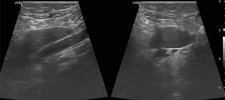
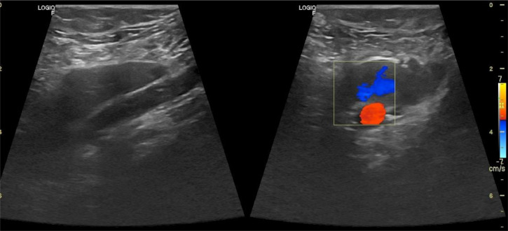
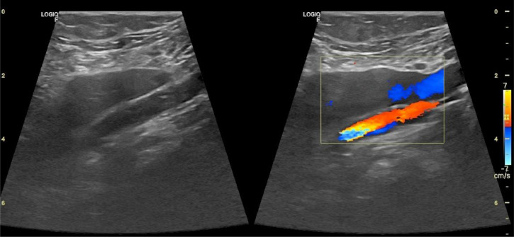
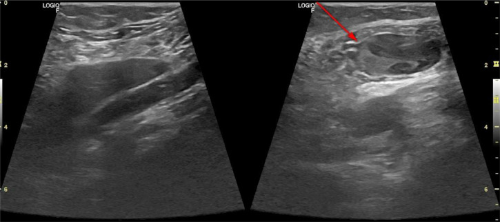
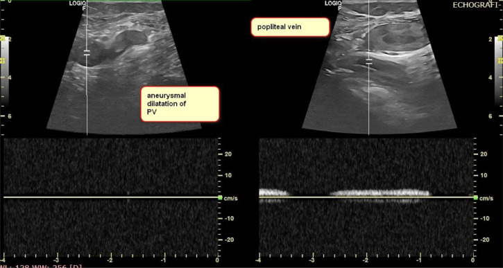
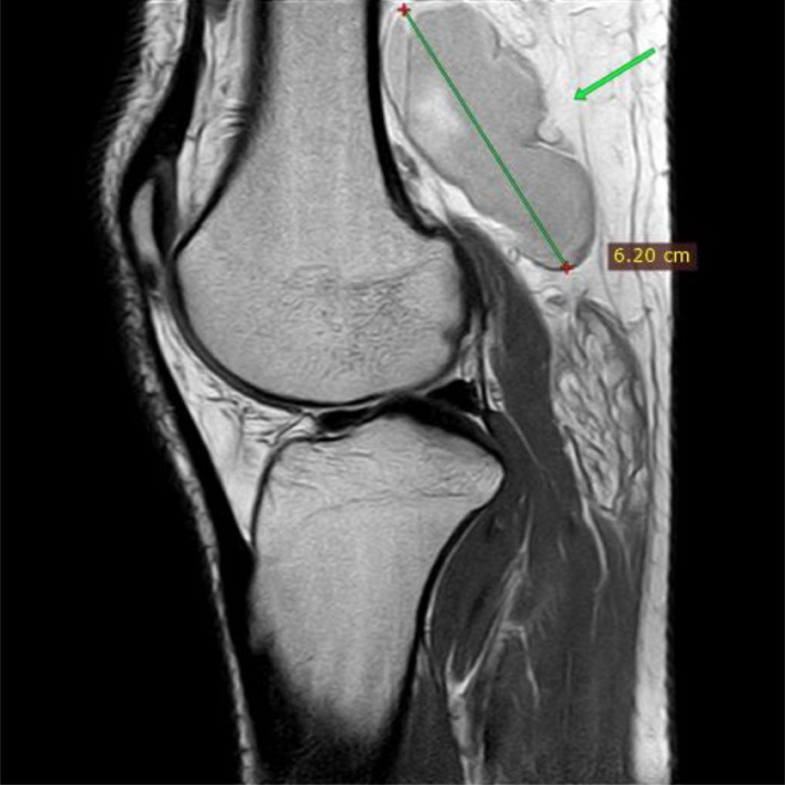
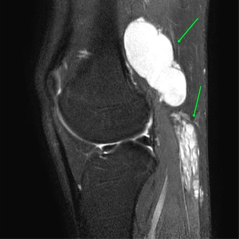
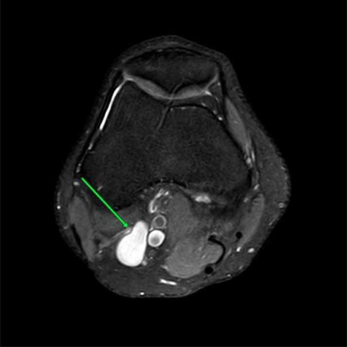
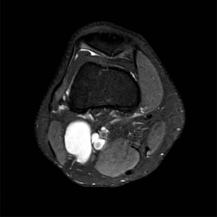
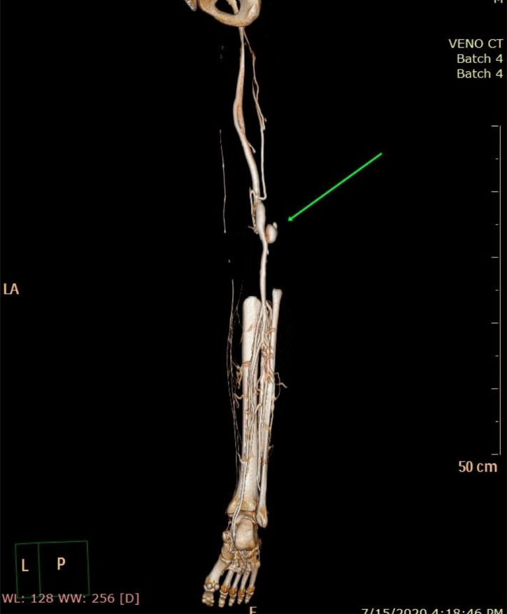
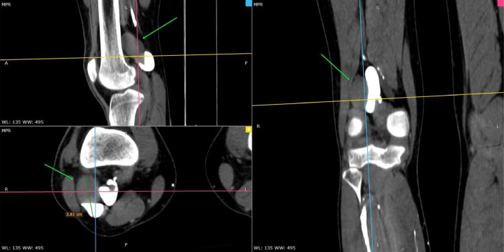
Primitive vein popliteal aneurysms are rare and potentially fatal vascular disorders. The most dangerous complications of popliteal vein aneurysms are thromboembolic events, mainly pulmonary embolisms, a life-threatening event that requires a timely diagnosis and prompt management. As a treatable cause of recurrent pulmonary embolisms, their actual incidence is believed to be underestimated. Herein, we present a case report of a popliteal vein aneurysm in a previously healthy16-year old male, presenting with a swelling behind his left knee that causes minimal discomfort while walking. When feasible, early surgical repair of both symptomatic and asymptomatic popliteal venous aneurysms is advised, since they are associated with an ill-defined possibility of pulmonary embolism and mortality, if left untreated.
Keywords: Asymptomatic; Duplex ultrasound; MRI angiography; Popliteal vein aneurysm; Pulmonary embolism.
© 2021 The Authors. Published by Elsevier Inc. on behalf of University of Washington.
Figures
References
-
- Maleti O, Lugli M, Collura M. Anéurysmes veineux poplités: experience personelle. Phlebology. 1997;50:53–59.
Publication types
LinkOut - more resources
Full Text Sources
Other Literature Sources
Research Materials
