Case Reports
. 2021 Apr 13;16(6):1463-1468.
doi: 10.1016/j.radcr.2021.03.010. eCollection 2021 Jun.

Cerebellar developmental venous anomaly with associated cavernoma causing a hemorrhage - a rare occurrence

Affiliations
Case Reports

Cerebellar developmental venous anomaly with associated cavernoma causing a hemorrhage - a rare occurrence

Omar Marzouk et al. Radiol Case Rep. .

Abstract

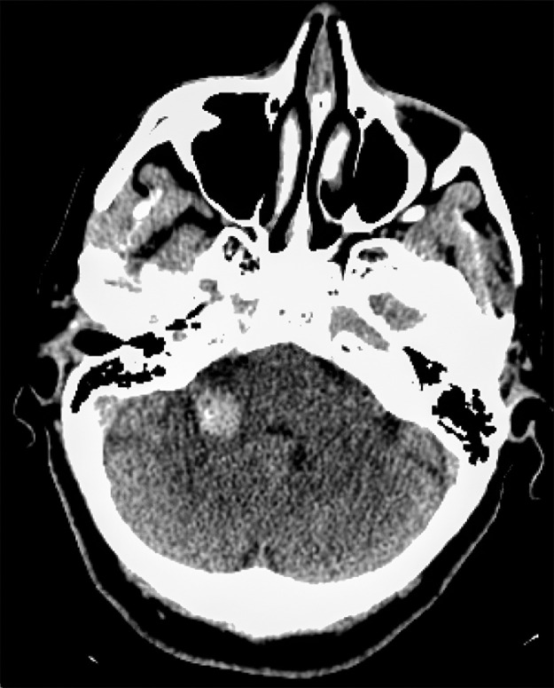
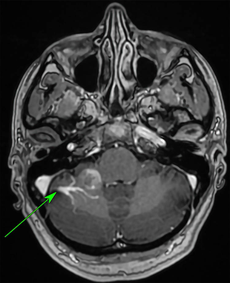
Developmental venous anomalies (DVAs) are variations in the transmedullary veins required for drainage of the brain. Normally, when occurring in isolation, DVAs are not clinically significant and are therefore usually a benign diagnosis. Thus, they are most often an incidental finding unless associated with an adjacent pathology. However, intracranial haemorrhage induced by a DVA alone can rarely occur and has been scarcely reported. In this case report we discuss a 58-year-old woman who presented with signs and symptoms of a cerebellar syndrome. Following a non-contrast CT, a CT angiogram and MRI contrast scan of the brain, she was found to have a cerebellar DVA and an intracranial haemorrhage. Subsequent imaging 3 months later with CT and MRI redemonstrated additional evidence of a cavernoma. The patient was managed conservatively.

Keywords: Arteriovenous malformation; Cavernoma; Cerebellar developmental venous anomaly; Infratentorial developmental venous anomaly; Intracranial haemorrhage; Venous angioma.


Figures

Fig 1 –
Fig. 1
Axial CT Head: Initial non enhanced CT showing acute intracerebral hemorrhage in the right cerebellar peduncle.
Fig 2
Fig. 2
Axial CT Angiogram: The DVA is again well demonstrated. A connecting vein is seen (green arrow) to a dural. (color version of figure is available online.)
Fig 3 –
Fig. 3
Coronal CT Angiogram: Maximum intensity projection reconstruction demonstrating the classically described palm tree DVA is again well demonstrated.
Fig 4 –
Fig. 4
Sagittal CT Angiogram: Maximum intensity projection reconstruction demonstrating the classically described caput.
Fig 5 –
Fig. 5
T1w volume Bravo pre contrast: A branching signal void is seen in keeping with DVA (green arrow). A focus of high signal. (color version of figure is available online.)
Fig 6 –
Fig 6
T1w volume Bravo post contrast: The previously seen branching venous anomaly is well demonstrated with avid contrast.
Fig 7 –
Fig. 7
SWI sequence: The DVA is again well seen. Extensive blooming artefact is seen associated with the recent hemorrhage.
Fig 8 –
Fig 8
Repeat axial CT head: resolution of the previous hemorrhage with a focus of calcification in the middle cerebellar.
Fig 9 –
Fig. 9
Repeat T1w volume: The DVA is again demonstrated with a nearby low signal focus. No areas of high signal to indicate.
Fig 10 –
Fig. 10
Repeat SWI: Site of cavernoma indicated by green arrow. A blooming artefact with hypointense limb demonstrating.

References

    1. Mooney MA, Zabramski JM. Developmental venous anomalies. Handb Clin Neurol. 2017:279–282. doi: 10.1016/b978-0-444-63640-9.00026-6. - DOI - PubMed
    1. Lasjaunias P, Burrows P, Planet C. Developmental venous anomalies (DVA): the so-called venous angioma. Neurosurg Rev. 1986;9(3):233–242. doi: 10.1007/bf01743138. - DOI - PubMed
    1. Boukobza M, Enjolras O, Guichard J P, Gelbert F, Herbreteau D, Reizine D. Cerebral developmental venous anomalies associated with head and neck venous malformations. AJNR Am J Neuroradiol. 1996;17(5):987–994. http://www.ajnr.org/content/17/5/987 - PMC - PubMed
    1. Pereira VM, Geibprasert S, Krings T, Aurboonyawat T, Ozanne A, Toulgoat F. Pathomechanisms of symptomatic developmental venous anomalies. Stroke. 2008;39(12):3201–3215. doi: 10.1161/STROKEAHA.108.521799. - DOI - PubMed
    1. Campeau NG, Lane JI. De novo development of a lesion with the appearance of a cavernous malformation adjacent to an existing developmental venous anomaly. Am J Neuroradiol. 2005;26(1):156–159. - PMC - PubMed

Publication types

LinkOut - more resources