

Cutaneous and hepatic vascular lesions due to a recurrent somatic GJA4 mutation reveal a pathway for vascular malformation
- PMID: 33912852
- PMCID: PMC8078848
- DOI: 10.1016/j.xhgg.2021.100028
Cutaneous and hepatic vascular lesions due to a recurrent somatic GJA4 mutation reveal a pathway for vascular malformation
Erratum in
-
Erratum: Cutaneous and hepatic vascular lesions due to a recurrent somatic GJA4 mutation reveal a pathway for vascular malformation.HGG Adv. 2021 Oct 22;3(1):100061. doi: 10.1016/j.xhgg.2021.100061. eCollection 2022 Jan 13. HGG Adv. 2021. PMID: 35047851 Free PMC article.
Abstract
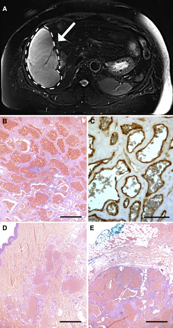
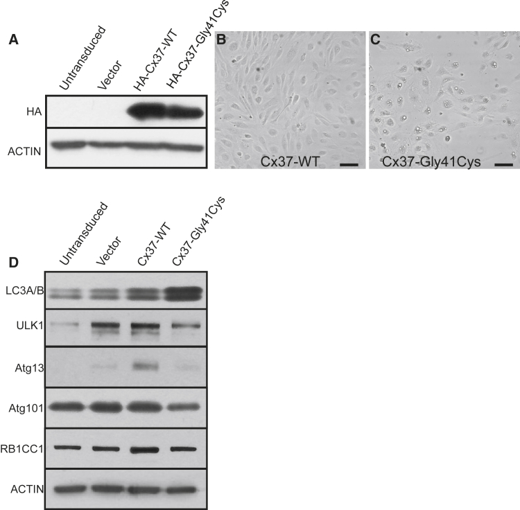
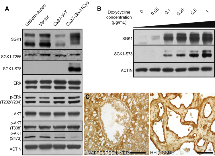
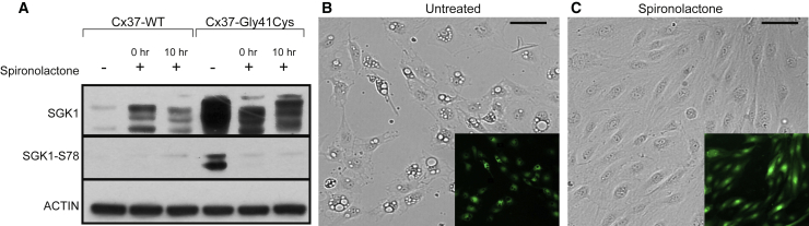
The term "cavernous hemangioma" has been used to describe vascular anomalies with histology featuring dilated vascular spaces, vessel walls consisting mainly of fibrous stromal bands lined by a layer of flattened endothelial cells, and an irregular outer rim of interrupted smooth muscle cells. Hepatic hemangiomas (HHs) and cutaneous venous malformations (VMs) share this histologic pattern, and we examined lesions in both tissues to identify genetic drivers. Paired whole-exome sequencing (WES) of lesional tissue and normal liver in HH subjects revealed a recurrent GJA4 c.121G>T (p.Gly41Cys) somatic mutation in four of five unrelated individuals, and targeted sequencing in paired tissue from 9 additional HH individuals identified the same mutation in 8. In cutaneous lesions, paired targeted sequencing in 5 VMs and normal epidermis found the same GJA4 c.121G>T (p.Gly41Cys) somatic mutation in three. GJA4 encodes gap junction protein alpha 4, also called connexin 37 (Cx37), and the p.Gly41Cys mutation falls within the first transmembrane domain at a residue highly conserved among vertebrates. We interrogated the impact of the Cx37 mutant via lentiviral transduction of primary human endothelial cells. We found that the mutant induced changes in cell morphology and activated serum/glucocorticoid-regulated kinase 1 (SGK1), a serine/threonine kinase known to regulate cell proliferation and apoptosis, via non-canonical activation. Treatment with spironolactone, an inhibitor of angiogenesis, suppressed mutant SGK1 activation and reversed changes in cell morphology. These findings identify a recurrent somatic GJA4 c.121G>T mutation as a driver of hepatic and cutaneous VMs, revealing a new pathway for vascular anomalies, with spironolactone a potential pathogenesis-based therapy.
Conflict of interest statement
Declaration of interests The authors declare no competing interests.
Figures
Similar articles
-
Somatic GJA4 mutation in intracranial extra-axial cavernous hemangiomas.Stroke Vasc Neurol. 2023 Dec 29;8(6):453-462. doi: 10.1136/svn-2022-002227. Stroke Vasc Neurol. 2023. PMID: 37072338 Free PMC article.
-
Somatic GJA4 gain-of-function mutation in orbital cavernous venous malformations.Angiogenesis. 2023 Feb;26(1):37-52. doi: 10.1007/s10456-022-09846-5. Epub 2022 Jul 29. Angiogenesis. 2023. PMID: 35902510 Free PMC article.
-
The dural angioleiomyoma harbors frequent GJA4 mutation and a distinct DNA methylation profile.Acta Neuropathol Commun. 2022 May 31;10(1):81. doi: 10.1186/s40478-022-01384-x. Acta Neuropathol Commun. 2022. PMID: 35642047 Free PMC article.
-
A clinicopathological reappraisal of orbital vascular malformations and distinctive GJA4 mutation in cavernous venous malformations.Hum Pathol. 2022 Dec;130:79-87. doi: 10.1016/j.humpath.2022.10.002. Epub 2022 Oct 6. Hum Pathol. 2022. PMID: 36209871 Review.
-
Genetic landscape of common venous malformations in the head and neck.J Vasc Surg Venous Lymphat Disord. 2021 Jul;9(4):1007-1016.e7. doi: 10.1016/j.jvsv.2020.11.016. Epub 2020 Nov 26. J Vasc Surg Venous Lymphat Disord. 2021. PMID: 33248299 Review.
Cited by
-
Vascular and Lymphatic Malformations: Perspectives From Human and Vertebrate Studies.Circ Res. 2021 Jun 25;129(1):131-135. doi: 10.1161/CIRCRESAHA.121.319587. Epub 2021 Jun 24. Circ Res. 2021. PMID: 34166069 Free PMC article.
-
Somatic GJA4 mutation in intracranial extra-axial cavernous hemangiomas.Stroke Vasc Neurol. 2023 Dec 29;8(6):453-462. doi: 10.1136/svn-2022-002227. Stroke Vasc Neurol. 2023. PMID: 37072338 Free PMC article.
-
Somatic GJA4 gain-of-function mutation in orbital cavernous venous malformations.Angiogenesis. 2023 Feb;26(1):37-52. doi: 10.1007/s10456-022-09846-5. Epub 2022 Jul 29. Angiogenesis. 2023. PMID: 35902510 Free PMC article.
-
Mesenchymal non-meningothelial tumors of the central nervous system: a literature review and diagnostic update of novelties and emerging entities.Acta Neuropathol Commun. 2023 Feb 3;11(1):22. doi: 10.1186/s40478-023-01522-z. Acta Neuropathol Commun. 2023. PMID: 36737790 Free PMC article. Review.
-
The dural angioleiomyoma harbors frequent GJA4 mutation and a distinct DNA methylation profile.Acta Neuropathol Commun. 2022 May 31;10(1):81. doi: 10.1186/s40478-022-01384-x. Acta Neuropathol Commun. 2022. PMID: 35642047 Free PMC article.
References
-
- Funk T., Lim Y., Kulungowski A.M., Prok L., Crombleholme T.M., Choate K., Bruckner A.L. Symptomatic Congenital Hemangioma and Congenital Hemangiomatosis Associated With a Somatic Activating Mutation in GNA11. JAMA Dermatol. 2016;152:1015–1020. - PubMed
-
- Groesser L., Peterhof E., Evert M., Landthaler M., Berneburg M., Hafner C. BRAF and RAS Mutations in Sporadic and Secondary Pyogenic Granuloma. J. Invest. Dermatol. 2016;136:481–486. - PubMed
-
- Lim Y.H., Bacchiocchi A., Qiu J., Straub R., Bruckner A., Bercovitch L., Narayan D., McNiff J., Ko C., Robinson-Bostom L., et al. Yale Center for Mendelian Genomics GNA14 Somatic Mutation Causes Congenital and Sporadic Vascular Tumors by MAPK Activation. Am. J. Hum. Genet. 2016;99:443–450. - PMC - PubMed
Grants and funding
LinkOut - more resources
Full Text Sources
Other Literature Sources
Molecular Biology Databases
Miscellaneous
