Case Reports
. 2021 Jun 16;106(7):1867-1881.
doi: 10.1210/clinem/dgab283.

A Novel SLC5A5 Variant Reveals the Crucial Role of Kinesin Light Chain 2 in Thyroid Hormonogenesis

Affiliations
Case Reports

A Novel SLC5A5 Variant Reveals the Crucial Role of Kinesin Light Chain 2 in Thyroid Hormonogenesis

Mariano Martín et al. J Clin Endocrinol Metab. .

Abstract

Context: Iodide transport defect (ITD) (Online Mendelian Inheritance in Man No. 274400) is an uncommon cause of dyshormonogenic congenital hypothyroidism due to loss-of-function variants in the SLC5A5 gene, which encodes the sodium/iodide symporter (NIS), causing deficient iodide accumulation in thyroid follicular cells.

Objective: This work aims to determine the molecular basis of a patient's ITD clinical phenotype.

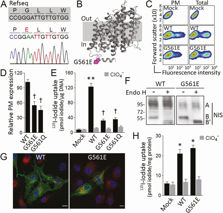
Methods: The propositus was diagnosed with dyshormonogenic congenital hypothyroidism with minimal 99mTc-pertechnetate accumulation in a eutopic thyroid gland. The propositus SLC5A5 gene was sequenced. Functional in vitro characterization of the novel NIS variant was performed.

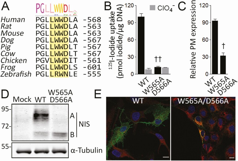
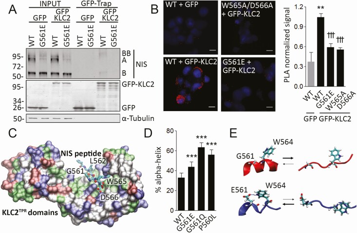
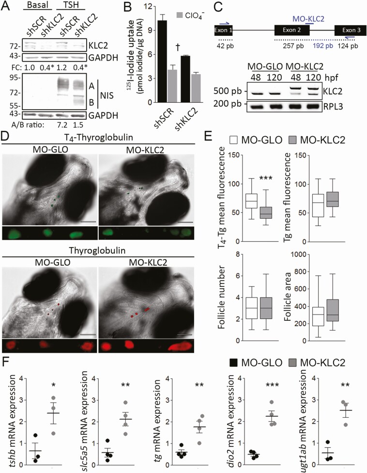
Results: Sanger sequencing revealed a novel homozygous missense p.G561E NIS variant. Mechanistically, the G561E substitution reduces iodide uptake, because targeting of G561E NIS to the plasma membrane is reduced. Biochemical analyses revealed that G561E impairs the recognition of an adjacent tryptophan-acidic motif by the kinesin-1 subunit kinesin light chain 2 (KLC2), interfering with NIS maturation beyond the endoplasmic reticulum, and reducing iodide accumulation. Structural bioinformatic analysis suggests that G561E shifts the equilibrium of the unstructured tryptophan-acidic motif toward a more structured conformation unrecognizable to KLC2. Consistently, knockdown of Klc2 causes defective NIS maturation and consequently decreases iodide accumulation in rat thyroid cells. Morpholino knockdown of klc2 reduces thyroid hormone synthesis in zebrafish larvae leading to a hypothyroid state as revealed by expression profiling of key genes related to the hypothalamic-pituitary-thyroid axis.

Conclusion: We report a novel NIS pathogenic variant associated with dyshormonogenic congenital hypothyroidism. Detailed molecular characterization of G561E NIS uncovered the significance of KLC2 in thyroid physiology.

Keywords: dyshormonogenic congenital hypothyroidism; impaired transport to the plasma membrane; iodide transport defect; kinesin light chain 2; sodium/iodide symporter; tryptophan-acidic motif.


Figures

Figure 1.
Figure 1.
G561E severely reduces sodium/iodide symporter (NIS) plasma membrane expression. A, Chromatogram showing a 15-bp fragment of NIS-coding exon 14 (nucleotides + 1.678 to + 1.692). The reference sequence (Refseq) is indicated in gray; the variant p.G561E is indicated in red. B, NIS homology model highlighting the cytosol-facing residue E561 in magenta. C, Representative flow cytometry analysis (pseudo-color density plot, forward scatter vs fluorescence intensity) of NIS expression at the plasma membrane (PM) and whole-cell (total) in cells permanently expressing vector (Mock), wild-type (WT) or G561E NIS. D, Relative WT or mutant NIS expression at the plasma membrane assessed by flow cytometry under nonpermeabilized conditions. Data are mean ± SEM. †P < .05 vs WT NIS-transfected cells (Kruskal-Wallis, Dunn post hoc test). E, Steady-state iodide uptake in cells permanently expressing WT, G561E, or G561Q NIS. Results are expressed in pmol iodide/μg DNA ± SEM. **P < .01 vs mock-transfected cells, †P < .05 vs WT NIS-transfected cells (Kruskal-Wallis, Dunn post hoc test). F, Endo-β-acetylglucosaminidase H (Endo H) treatment of whole-cell lysates from cells expressing WT or G561E NIS. Labels indicate the relative electrophoretic mobilities of the corresponding NIS polypeptides (A: Endo H resistant, fully glycosylated; B and B’: Endo H sensitive, partially glycosylated before and after Endo H digestion) depending on glycosylation status. G, Confocal microscopy analysis of permeabilized cells expressing WT or G561E NIS. Nuclei were stained with 4′, 6-diamidino-2-phenylindole (DAPI) (blue). The overlay of green (NIS) and red (KDEL) channels is shown. Scale bar: 10 μm. H, Iodide uptake in membrane vesicles prepared from cells expressing empty vector (Mock), WT, or G561E NIS. Results are expressed as pmol iodide/mg protein ± SEM. *P < .05 vs mock-transfected cells (Kruskal-Wallis, Dunn post hoc test).
Figure 2.
Figure 2.
A conserved tryptophan-acidic motif is required for sodium/iodide symporter (NIS) endoplasmic reticulum (ER) exit. A, Multiple NIS sequences alignment. The conserved tryptophan-acidic motif in the NIS carboxy-terminus is highlighted. Sequence logo represents degree of conservation. B, Steady-state iodide uptake in cells permanently expressing wild-type (WT) or W565A/D566A NIS. Results are expressed in pmol iodide/μg DNA ± SEM. ††P < .01 vs WT NIS-transfected cells (Kruskal-Wallis, Dunn post hoc test). C, Relative WT or mutant NIS expression at the plasma membrane assessed by flow cytometry. Data are mean ± SEM. †P < .05 vs WT NIS-transfected cells (Mann-Whitney U test). D, Immunoblot analysis of whole-cell lysates from cells expressing WT or W565A/D566A NIS. Labels indicate the relative electrophoretic mobilities of the corresponding NIS polypeptides (A: Fully glycosylated; B: partially glycosylated) depending on glycosylation status. α-tubulin is shown as a loading control. E, Confocal microscopy analysis of permeabilized cells expressing WT or W565A/D566A NIS. Nuclei were stained with ′, 6-diamidino-2-phenylindole (DAPI) (blue). The overlay of green (NIS) and red (KDEL) channels is shown. Scale bar: 10 μm.
Figure 3.
Figure 3.
G561E interferes with the interaction between sodium/iodide symporter (NIS) and kinesin-1 light chain 2. A, Immunoprecipitation of green fluorescent protein (GFP) with GFP-Trap technology using whole-cell lysates from type II Madin–Darby canine kidney (MDCK-II) cells permanently expressing wild-type (WT) or G561E NIS and transiently transfected with GFP or GFP-KLC2. Labels indicate the relative electrophoretic mobilities of the corresponding GFP-Trap pulled-down NIS polypeptides (A: Fully glycosylated; B and BB: monomer and dimer of partially glycosylated) depending on glycosylation status. Labels on the right side of the blot indicate the relative electrophoretic mobilities of the corresponding NIS polypeptides. Undetectable α-tubulin protein levels demonstrate the absence of cytoplasmic contamination in GFP-Trap pull-downs. B, Proximity ligation assay (PLA) in human embryonic kidney (HEK)-293T cells cotransfected with WT, W565A/D566A, or G561E NIS along with GFP or GFP-KLC2. **P < .01 vs WT + GFP, †††P < .001 vs WT + GFP-KLC2 (Kruskal-Wallis, Dunn post hoc test). Scale bar: 10 μm. C, Tryptophan-acidic motif-containing NIS octapeptide P560-L567 bound to the KLC2 tetratricopeptide (KLC2TPR) domains represented as a molecular surface colored by residue type. D, Percentage of α-helix structure adopted by WT, G561E, G561Q, and P560L NIS octapeptides P560-L567 during the molecular dynamic simulation. ***P < .001 (analysis of variance, Bonferroni post hoc test). E, Structural equilibrium of WT (red) or G561E (blue) octapeptide P560-L567. A putative hydrogen bond between residues E561 and W564 is indicated.
Figure 4.
Figure 4.
KLC2 knockdown reduced thyroid hormonogenesis due to defective sodium/iodide symporter (NIS)-mediated iodide accumulation. A, Immunoblot analysis of whole-cell lysates from scrambled (SCR) or Klc2 short hairpin RNA (shRNA)-transfected PCCl3 cells in response to thyrotropin (TSH) treatment for 48 hours. Labels indicate the relative electrophoretic mobilities of the corresponding NIS polypeptides (A: Fully glycosylated; B: partially glycosylated) depending on glycosylation status. Glyceraldehyde-3-phosphate dehydrogenase (GAPDH) expression was used as loading control. *P < .05 vs SCR shRNA-transfected cells (Mann-Whitney U test). Fold change (FC) represents the mean from 3 independent experiments. B, Steady-state iodide uptake in SCR or Klc2 shRNA-transfected PCCl3. Results are expressed in pmol iodide/μg DNA ± SEM. †P < .05 vs SCR shRNA-transfected cells (Kruskal-Wallis, Dunn post hoc test). C, Partial schematic representation of the klc2 gene. Anti-klc2 morpholino (MO-KLC2) and primer hybridization’s sites are indicated. Reverse transcriptase–polymerase chain reaction assessing KLC intron 2 inclusion in control (MO-GLO) or anti-klc2 (MO-KLC2) morpholino-microinjected larvae at 48 and 120 hpf. The expression of rpl-3 was used as loading control. D, Representative whole-mount immunofluorescence assessing thyroglobulin (Tg)-bound thyroxine (T4-Tg) and Tg in embryos microinjected with MO-GLO or MO-KLC2 at 120 hpf. Scale bar: 100 μm. Close-up shows representative selection of immunostainings (magnified ventral views of thyroid region). E, Quantification of mean fluorescence intensity of T4-Tg and Tg, and number and cross-section area (μm2) of T4-Tg immunoreactive follicles in embryos microinjected with MO-GLO or MO-KLC2 at 120 hpf. ***P < .001 vs MO-GLO–microinjected larvae (Mann-Whitney U test). The number of larvae analyzed per condition was 35. F, Relative whole-body messenger RNA (mRNA) expression levels of genes related to the hypothalamic-pituitary-thyroid axis in MO-GLO– or MO-KLC2–microinjected embryos at 120 hpf. *P < .05, **P < .01 vs MO-GLO–microinjected larvae (Mann-Whitney U test). The number of larvae analyzed per replicate was 30.

References

    1. Szinnai G. Genetics of normal and abnormal thyroid development in humans. Best Pract Res Clin Endocrinol Metab. 2014;28(2):133-150. - PubMed
    1. Fujiwara H, Tatsumi K, Miki K, et al. . Congenital hypothyroidism caused by a mutation in the Na+/I– symporter. Nat Genet. 1997;16(2):124-125. - PubMed
    1. Matsuda A, Kosugi S. A homozygous missense mutation of the sodium/iodide symporter gene causing iodide transport defect. J Clin Endocrinol Metab. 1997;82(12):3966-3971. - PubMed
    1. Nicola JP, Carrasco N, Masini-Repiso AM. Chapter one—Dietary I– absorption: expression and regulation of the Na+/I– symporter in the intestine. Vitam Horm. 2015;98:1-31. - PubMed
    1. Martín M, Geysels RC, Peyret V, Bernal Barquero CE, Masini-Repiso AM, Nicola JP.. Implications of Na+/I– symporter transport to the plasma membrane for thyroid hormonogenesis and radioiodide therapy. J Endoc Soc. 2019;3(1):222-234. - PMC - PubMed

Publication types