. 2021 May 1;148(9):dev197111.
doi: 10.1242/dev.197111. Epub 2021 Apr 26.

Testicular germ cell tumors arise in the absence of sex-specific differentiation

Affiliations

Testicular germ cell tumors arise in the absence of sex-specific differentiation

Nicholas J Webster et al. Development. .

Abstract

In response to signals from the embryonic testis, the germ cell intrinsic factor NANOS2 coordinates a transcriptional program necessary for the differentiation of pluripotent-like primordial germ cells toward a unipotent spermatogonial stem cell fate. Emerging evidence indicates that genetic risk factors contribute to testicular germ cell tumor initiation by disrupting sex-specific differentiation. Here, using the 129.MOLF-Chr19 mouse model of testicular teratomas and a NANOS2 reporter allele, we report that the developmental phenotypes required for tumorigenesis, including failure to enter mitotic arrest, retention of pluripotency and delayed sex-specific differentiation, were exclusive to a subpopulation of germ cells failing to express NANOS2. Single-cell RNA sequencing revealed that embryonic day 15.5 NANOS2-deficient germ cells and embryonal carcinoma cells developed a transcriptional profile enriched for MYC signaling, NODAL signaling and primed pluripotency. Moreover, lineage-tracing experiments demonstrated that embryonal carcinoma cells arose exclusively from germ cells failing to express NANOS2. Our results indicate that NANOS2 is the nexus through which several genetic risk factors influence tumor susceptibility. We propose that, in the absence of sex specification, signals native to the developing testis drive germ cell transformation.

Keywords: Nanos2; Germ cells; MYC; NODAL; TGCTs; Teratomas.


Conflict of interest statement

Competing interests The authors declare no competing or financial interests.

Figures

Fig. 1.
Fig. 1.
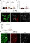
Tumor susceptible germ cells are delayed in expressing NANOS2RFP. (A) Germ cells from low TGCT risk 129 and high TGCT risk M19 embryos were FACS enriched using OCT4::GFP and Nanos2RFP, and the proportion of GFP-positive, Nanos2RFP-negative germ cells reported (n=3-13). (B) Proportion of E15.5 Nanos2RFP-negative, GFP-positive germ cells in 129 and M19 mice by manual counting of confocal images (n=6 and 24). (C) Representative image of testis from E15.5 M19.OCT4-GFP, Nanos2RFP mice sectioned and immunostained for NANOG to identify foci of embryonal carcinoma cells (yellow dashed outline). (D) GFP-positive, Nanos2RFP-negative and GFP-positive, Nanos2RFP-positive germ cells from M19 mice were FACS-enriched from E13.5 to E15.5 and analyzed by QPCR for Nanos2 (n=3-16). Data are mean±s.e.m. with individual data points indicated. *P≤0.05, **P≤0.01, SC, somatic cell.
Fig. 2.
Fig. 2.
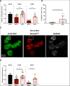
Nanos2RFP-deficient germ cells downregulate markers of PGC identity and delay expression of Dnmt3l, a marker of male germ cell sex specification. FACS-enriched OCT4::GFP-positive, Nanos2RFP-negative and OCT4::GFP-positive, Nanos2RFP-positive germ cells from M19 mice were analyzed by QPCR for (A) Prdm1 (n=3-5) and (B) Dnmt3l (n=3-16). (C) Testes of E15.5 M19 mice were sectioned and immunostained for DNMT3L. Germ cells were identified by GFP expression and the proportions of germ cells expressing DNMT3L and RFP reported (n=6). (D) Representative image of DNMT3L staining (RFP-negative, DNMT3L-negative germ cells outlined in white). ****P≤0.0001. Data are mean±s.e.m. with individual data points indicated.
Fig. 3.
Fig. 3.
NANOS2RFP-deficient germ cells are enriched for proliferation. (A) FACS-enriched OCT4::GFP-positive, Nanos2RFP-negative and OCT4::GFP-positive, Nanos2RFP-positive germ cells from M19 mice were analyzed by QPCR for Ccnd1 (n=3-16). (B) Testes of E15.5 M19 mice were sectioned and immunostained for CCND1. Germ cells were identified by OCT4::GFP expression and the proportions of germ cells expressing CCND1 and RFP reported (n=6). (C) Representative image of CCND1 staining (RFP-negative, CCND1-positive germ cells outlined in white). (D) Testes of E15.5 M19 mice were sectioned and immunostained for KI67. Germ cells were identified by GFP expression and proportions of germ cells expressing KI67 and RFP reported (n=6). (E) Representative image of KI67 staining (Nanos2RFP-negative, KI67-positive germ cells outlined in white). *P≤0.05, **P≤0.01, ****P≤0.0001. Data are mean±s.e.m. with individual data points indicated.
Fig. 4.
Fig. 4.
NANOS2RFP-deficient germ cells are enriched for pluripotency factors. (A) FACS-enriched OCT4::GFP-positive, Nanos2RFP-negative and OCT4::GFP-positive, Nanos2RFP-positive germ cells from M19 mice were analyzed by QPCR for Nanog (n=3-16). (B) Testes of E15.5 M19 mice were sectioned and immunostained for NANOG. Germ cells were identified by GFP expression and proportions of germ cells expressing NANOG and RFP reported (n=6). (C) Representative image of nuclear NANOG staining (Nanos2RFP-negative, NANOG-positive germ cells outlined in white). (D) QPCR analysis for Otx2. *P≤0.05, **P≤0.01, ****P≤0.0001. Data are mean±s.e.m. with individual data points indicated.
Fig. 5.
Fig. 5.
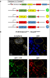
Embryonal carcinoma cells arise from precursors that do not initiate expression of NANOS2. (A) Following Cre-mediated recombination from the Nanos2Cre knock-in allele, Confetti allows the expression of GFP, YFP, RFP or CFP in any cell or daughter cell that expressed Nanos2 at any point in development. (B) Representative image of testis of a postnatal day 2 M19.Nanos2Cre/+ ; Rosa26Confetti/+ mouse. Sections were immunostained for E-cadherin to identify foci of embryonal carcinoma cells (outlined in yellow) and CFP (to amplify signal for imaging). GFP and YFP signals are combined due to technical restraints of the confocal imaging system used.
Fig. 6.
Fig. 6.
Nanos2RFP-negative germ cells and embryonal carcinoma cells cluster separately from somatic cells and Nanos2RFP-positive germ cells. OCT4::GFP, Nanos2RFP-positive and OCT4::GFP, Nanos2RFP-negative germ cells from were pooled from four M19 mice and were FACS enriched to be submitted for single-cell RNA sequencing. Transcriptomes were normalized and supervised clustering performed to yield 13 clusters. (A) Heatmap of a priori genes used as markers of pluripotent states, embryonic germ cell differentiation states, somatic cell identity and Nodal signaling were used to determine cell identities of each cluster. (B) List of identified cell types and composition of each cluster.
Fig. 7.
Fig. 7.
Embryonal carcinoma cells upregulate Myc signaling pathways. (A,B) Representative gene sets from Gene Set Enrichment Analysis of differential gene expression analysis performed comparing the average of (A) groups 4 and 5 (Nanos2RFP-negative, undifferentiated germ cells) to the average of groups 2 and 3 (Nanos2RFP-positive, differentiated germ cells) and (B) group 9 (containing embryonal carcinoma cells) to the average of groups 4 and 5 (RFP-negative, undifferentiated germ cells).
Fig. 8.
Fig. 8.
Model of tumorigenesis in mice. (A) During normal development, XY PGCs in mice expressing core pluripotency factors, such as NANOG, and primordial germ cell (PGC)-associated factors, such as PRDM1, migrate to and colonize the gonad. Following colonization, PGCs briefly express OTX2 followed by NANOS2 to enter sex-specific differentiation while simultaneously downregulating pluripotency. Concomitantly, TGFβ signaling present in the embryonic testis, primarily via activins and NODAL, promotes somatic cell growth and male germ cell development (not shown). (B) Tumorigenesis is proposed to occur due to a failure in the expression of male sex-specific genes, such as NANOS2, coinciding with the downregulation of PGC-associated factors. During this window of differentiation failure, expression of both core pluripotency and primed pluripotency factors is maintained and MYC signaling is upregulated. We propose that tumorigenesis occurs regardless of the cause of differentiation failure, potentially due to maintained response to TGFβ signaling, including defects accumulated during PGC migration that may affect differentiation post-colonization.

References

    1. Acampora, D., Di Giovannantonio, L. G. and Simeone, A. (2013). Otx2 is an intrinsic determinant of the embryonic stem cell state and is required for transition to a stable epiblast stem cell condition. Development 140, 43-55. 10.1242/dev.085290 - DOI - PubMed
    1. Adams, I. and Mclaren, A. (2002). Sexually dimorphic development of mouse primordial germ cells: Switching from oogenesis to spermatogenesis. Development 129, 1155-1164. - PubMed
    1. Ancelin, K., Lange, U. C., Hajkova, P., Schneider, R., Bannister, A. J., Kouzarides, T. and Surani, M. A. (2006). Blimp1 associates with Prmt5 and directs histone arginine methylation in mouse germ cells. Nat. Cell Biol. 8, 623-630. 10.1038/ncb1413 - DOI - PubMed
    1. Anderson, R., Fassler, R., Georges-Labouesse, E., Hynes, R. O., Bader, B. L., Kreidberg, J. A., Schaible, K., Heasman, J. and Wylie, C. (1999). Mouse primordial germ cells lacking beta1 integrins enter the germline but fail to migrate normally to the gonads. Development 126, 1655-1664. - PubMed
    1. Anderson, E. L., Baltus, A. E., Roepers-Gajadien, H. L., Hassold, T. J., de Rooij, D. G., van Pelt, A. M. M. and Page, D. C. (2008). Stra8 and its inducer, retinoic acid, regulate meiotic initiation in both spermatogenesis and oogenesis in mice. Proc. Natl Acad. Sci. USA 105, 14976. 10.1073/pnas.0807297105 - DOI - PMC - PubMed

Publication types

Supplementary concepts