. 2021 Jul;142(1):117-137.
doi: 10.1007/s00401-021-02313-3. Epub 2021 Apr 28.

LRP10 interacts with SORL1 in the intracellular vesicle trafficking pathway in non-neuronal brain cells and localises to Lewy bodies in Parkinson's disease and dementia with Lewy bodies

Affiliations

LRP10 interacts with SORL1 in the intracellular vesicle trafficking pathway in non-neuronal brain cells and localises to Lewy bodies in Parkinson's disease and dementia with Lewy bodies

Martyna M Grochowska et al. Acta Neuropathol. 2021 Jul.

Abstract

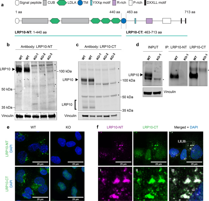
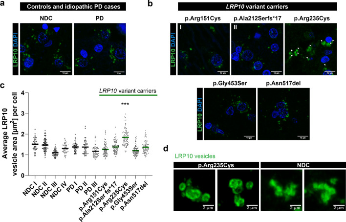
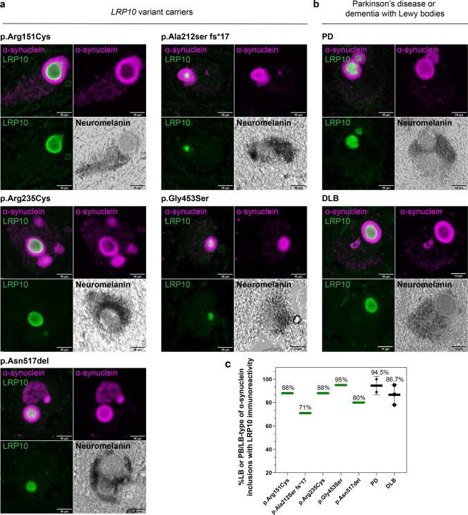
Loss-of-function variants in the low-density lipoprotein receptor-related protein 10 (LRP10) gene have been associated with autosomal-dominant Parkinson's disease (PD), PD dementia, and dementia with Lewy bodies (DLB). Moreover, LRP10 variants have been found in individuals diagnosed with progressive supranuclear palsy and amyotrophic lateral sclerosis. Despite this genetic evidence, little is known about the expression and function of LRP10 protein in the human brain under physiological or pathological conditions. To better understand how LRP10 variants lead to neurodegeneration, we first performed an in-depth characterisation of LRP10 expression in post-mortem brains and human-induced pluripotent stem cell (iPSC)-derived astrocytes and neurons from control subjects. In adult human brain, LRP10 is mainly expressed in astrocytes and neurovasculature but undetectable in neurons. Similarly, LRP10 is highly expressed in iPSC-derived astrocytes but cannot be observed in iPSC-derived neurons. In astrocytes, LRP10 is present at trans-Golgi network, plasma membrane, retromer, and early endosomes. Interestingly, LRP10 also partially co-localises and interacts with sortilin-related receptor 1 (SORL1). Furthermore, although LRP10 expression and localisation in the substantia nigra of most idiopathic PD and DLB patients and LRP10 variant carriers diagnosed with PD or DLB appeared unchanged compared to control subjects, significantly enlarged LRP10-positive vesicles were detected in a patient carrying the LRP10 p.Arg235Cys variant. Last, LRP10 was detected in Lewy bodies (LB) at late maturation stages in brains from idiopathic PD and DLB patients and in LRP10 variant carriers. In conclusion, high LRP10 expression in non-neuronal cells and undetectable levels in neurons of control subjects indicate that LRP10-mediated pathogenicity is initiated via cell non-autonomous mechanisms, potentially involving the interaction of LRP10 with SORL1 in vesicle trafficking pathways. Together with the specific pattern of LRP10 incorporation into mature LBs, these data support an important mechanistic role for disturbed vesicle trafficking and loss of LRP10 function in neurodegenerative diseases.

Keywords: Astrocytes; Dementia with Lewy bodies (DLB); LRP10; Lewy bodies; Parkinson’s disease (PD); Vesicle trafficking.


Conflict of interest statement

VB receives honoraria from the International Parkinson and Movement Disorder Society for serving as Chair of the MDS International Congress Program Committee 2019-2021; from Elsevier Ltd, for serving as co-Editor-in-Chief of Parkinsonism & Related Disorders; and from Springer, for serving as Section Editor of Current Neurology and Neuroscience Reports. WDJvdB was financially supported by grants from Amsterdam Neuroscience, Dutch Research council (ZonMW 70-73305-98-106; 70-73305-98-102; 40-46000-98-101), Stichting Parkinson Fonds (Insula 2014-2019), Alzheimer association (AARF-18-566459), MJ Fox foundation (17253) and Parkinson Association (2020-G01). WDJvdB performed contract research for Hoffmann-La Roche, Roche Tissue Diagnostics, Crossbeta Sciences, Lundbeck and received research consumables from Hoffmann-La Roche and Prothena. All other authors declare no competing interests.

Figures

Fig. 1
Fig. 1
Validation of LRP10 antibodies. a LRP10 protein structure. LRP10 antibodies were raised against the extracellular/luminal domain (LRP10-NT: 1–440 aa) or the intracellular/cytosolic domain (LRP10-CT: 463–713 aa). CUB, complement C1R/C1S, urchin EGF, BMP1; LDLA, low-density lipoprotein receptor class A; TM, transmembrane domain; R-rich, arginine-rich domain; P-rich, proline-rich domain; YXXφ motif of a tyrosine plus two other amino acids followed by an amino acid with a large bulky hydrophobic side chain; aa, amino acid. Western blots showing endogenous LRP10 in LRP10 WT and KO lines (black arrows) using extracellular (b) or intracellular (c) LRP10 antibody for detection. Whole cell lysates were obtained from HEK293T clones. Vinculin was used as a reference. asterisk, non-specific binding. d Immunoprecipitation of endogenous LRP10 from LRP10 WT and KO lines generated in HEK293T using both LRP10 antibodies. Input LRP10 was detected using LRP10-CT antibody. Immunoprecipitation with LRP10-NT, detection with LRP10-CT; immunoprecipitation with LRP10-CT, detection with LRP10-NT. Asterisk, non-specific binding. e Representative images of LRP10 (green) in LRP10 WT and KO lines from HuTu 80 cells. f Co-localisation of extracellular (magenta) and intracellular (green) LRP10 antibody in HuTu 80 cells. For e and f: maximum intensity projections. Scale bars, 25 µm. Nuclei were counterstained with DAPI (blue)
Fig. 2
Fig. 2
LRP10 is highly expressed in astrocytes and neurovasculature. a–e Representative images of LRP10 (green) protein expression at the cellular level in adult human midbrain. a Astrocytes were stained with S100β (magenta). b Dopaminergic neurons were stained with TH (magenta). c Microglia were stained with Iba1 (magenta). d Oligodendrocytes were stained with Quaking 7 (magenta), an RNA-binding protein that is highly specific for myelinating oligodendrocytes in the CNS. e Vascular smooth muscle cells were stained with α-SMA (magenta). Endothelial basement membrane is indicated between yellow, dashed lines. Yellow arrows indicate cells positive for either S100β, Iba1, or Quaking 7. f 3D surface rendering of LRP10 in astrocyte processes (GFAP, magenta) in adult human midbrain. LRP10 vesicle cluster around perinuclear region is indicated with red asterisks. Yellow arrows indicate partial co-localisation between LRP10 and GFAP. g RT-qPCR analysis of LRP10 and SNCA genes in human iPSC-derived progenitors, vmDAN, and astrocytes from a control line. Data represent relative normalized mRNA expression using primers designed against N-term and C-term of LRP10 or SNCA cDNA. CLK2, COPS5, RNF10 were used as reference genes. Error bars represent ± SEM (N = 3 biological replicates). h Representative images of LRP10 protein expression in human iPSC-derived dopaminergic neurons (TH) or astrocytes (S100β). Representative images from a minimum of three independent differentiations. a–f Representative images from a minimum of three brain sections derived from three non-demented individuals. All stainings were performed with LRP10-NT antibody. Nuclei were counterstained with DAPI (blue). Maximum intensity projections. Scale bars, 10 µm. vmDAN, ventral midbrain dopaminergic neurons
Fig. 3
Fig. 3
LRP10 is localised to vesicular structures and interacts with SORL1. a–d Deconvolved confocal images of LRP10 (green) co-localisation with various subcellular markers in 8 weeks old iPSC-derived astrocytes. a Early endosomes were detected using anti-EEA1 antibody (magenta). b The retromer complex was stained with anti-VPS35 antibody (magenta). c Trans-Golgi was stained with anti-TGN46 (magenta). d Lysosome-associated membrane protein 1 (LAMP-1) was used to detect lysosomal vesicles. a–d Zoomed images represent magnified views of boxed areas in the perinuclear region. Co-localisation with LRP10 is indicated by yellow arrowheads. Scale bars, 10 µm. e Quantification of LRP10 co-localisation with various subcellular markers. Thresholded Manders Overlap Coefficient is plotted on the y-axis. Per condition, 35 cells analysed. Error bars represent mean ± SD, one-way ANOVA with post hoc Tukey test; n.s., not significant; ***p < 0.001. f 11 weeks old human iPSC-derived astrocytes (SOX9+, magenta) were permeabilized and stained for intracellular LRP10 (green). For panels I and II: astrocytes were pre-incubated on ice to arrest endocytosis. Cell surface LRP10 molecules at the plasma membrane (PM) were labelled with LRP10-NT antibody. Scale bars, 10 µm. g, h Representative LRP10 (green) and SORL1 (magenta) co-localisation in iPSC-derived astrocytes (g) or in adult human midbrain (h). Images represent magnified views of boxed areas in the perinuclear region. Representative images from a minimum of three differentiations or three brain sections derived from three different non-demented individuals. Co-localisation LRP10 with SORL1 is indicated by yellow arrowheads. i–k HEK293T LRP10-KO or WT cells were transiently co-transfected with untagged full-length SORL1 only or together with N-terminally V5-tagged full-length LRP10. Total cell lysates (input) were subjected to immunoprecipitation with antibodies against SORL1 and LRP10. SORL1 antibody detected several bands (> 180 kDa) of overexpressed full-length SORL1 protein. Mouse IgG was used as a negative control. Immune complexes were resolved by SDS-PAGE, followed by Western blotting. a–d, f–h All stainings were performed with LRP10-NT antibody. Nuclei were counterstained with DAPI (blue). Maximum intensity projections
Fig. 4
Fig. 4
Enlarged LRP10-positive vesicles detected in brain of the LRP10 p.Arg235Cys variant carrier. a, b Representative images of LRP10 vesicle morphology from NDC (N = 4 individuals), PD (N = 3 individuals) (a), and five patients carrying LRP10 pathogenic variants (b). Enlarged and clustered LRP10 vesicles were seen in the midbrain of the PDD patient carrying p.Arg235Cys variant (white arrowheads). Representative images from a minimum of three brain sections. Nuclei were counterstained with DAPI (blue). Maximum intensity projections. All stainings were performed with the LRP10-NT antibody. Scale bars, 10 µm. c Quantification of surface area (µm2) of LRP10-postive vesicles in controls, idiopathic PD and LRP10 variants carriers. Each value in the scatter dot plots represents the average vesicle size/cell. Kruskal–Wallis with Dunn multiple comparison post hoc; ***p < 0.001. N equals the number of cells analysed per brain specimen. NNDC I = 108, NNDC II = 157, NNDC III = 112, NNDC IV = 58, NPD I = 112, NPD II = 96, NPD III = 80, Np.Arg151Cys = 87, Np.Ala212Ser fs*17 = 89, Np.Arg235Cys = 83, Np.Gly453Ser = 85, Np.Asn517del = 74. d High-resolution, re-scan confocal images of LRP10 vesicles reveal enlarged donut-like morphology in the PDD patient carrying p.Arg235Cys variant when compared to a non-demented control, scale bars 2 µm
Fig. 5
Fig. 5
LRP10 immunoreactivity in the core of nigral Lewy bodies. a, b Double labelling of α-synuclein (SYN-1, magenta) with LRP10 (green) antibody recognizing the intracellular domain. LRP10 immunoreactivity is present in the core of a LB in neuromelanin-positive dopaminergic neurons of SNpc (bright-field images). Representative confocal images of nigral LBs in patients carrying LRP10 mutations (a), or idiopathic PD and DLB (b). Maximum intensity projections. Scale bars, 10 µm. c Quantification of percentages of LRP10 immunoreactivity in more mature PB/LB-type or LB-type of α-synuclein inclusions. All inclusions analysed were found in neuromelanin-containing dopaminergic neurons of substantia nigra. N = 20 LB per individual; NDLB = 3 individuals (60 LB); NPD = 2 individuals (40 LB). Error bars represent mean ± SD. LB, Lewy body
Fig. 6
Fig. 6
Incorporation of LRP10 into α-synuclein inclusions during LB formation. a. Representative confocal images of morphological spectrum of α-synuclein (SYN-1, magenta) inclusion formation (steps 1–6). LRP10 (green) incorporation is present in more mature PB/LB or LB-type of α-synuclein inclusions. 1. Intracytoplasmic, punctuate α-synuclein. 2. Intracytoplasmic, punctuate α-synuclein with an aggregation centre (arrow). 3. PB-type: less compact, unstructured α-synuclein inclusion. Arrow heads and zoomed area indicate LRP10 vesicles. 4. Intracytoplasmic α-synuclein network containing PB (asterisk) and more compact LB-type inclusions (arrow). 5. Intracytoplasmic α-synuclein network with LB-type inclusion. 6. Mature LB. Maximum intensity projections. Scale bars, 10 µm. Representative images from a total of 247 nigral α-synuclein inclusions imaged. All inclusions analyzed were found in neuromelanin-containing dopaminergic neurons of substantia nigra. b Percentages of LRP10 immunoreactivity in different patterns of α-synuclein inclusions. Ntotal = 247; Npunctuate = 51; NPB-type = 44; NPB/LB-type or LB-type = 152. Kruskal–Wallis with Dunn multiple comparisons test; n.s., not significant; ***p < 0.001. LB, Lewy body. PB, pale body

References

    1. Aarsland D, Kvaløy JT, Andersen K, Larsen JP, Tang MX, Lolk A, et al. The effect of age of onset of PD on risk of dementia. J Neurol. 2007;254:38–45. - PubMed
    1. Abbott NJ, Rönnbäck L, Hansson E. Astrocyte-endothelial interactions at the blood-brain barrier. Nat Rev Neurosci. 2006;7:41–53. - PubMed
    1. Abeliovich A, Gitler AD. Defects in trafficking bridge Parkinson's disease pathology and genetics. Nature. 2016;539:207–216. doi: 10.1038/nature20414. - DOI - PubMed
    1. Allen NJ, Eroglu C. Cell biology of astrocyte-synapse interactions. Neuron. 2017;96:697–708. - PMC - PubMed
    1. Andersen OM, Reiche J, Schmidt V, Gotthardt M, Spoelgen R, Behlke J, et al. Neuronal sorting protein-related receptor sorLA/LR11 regulates processing of the amyloid precursor protein. Proc Natl Acad Sci USA. 2005;102:13461–13466. - PMC - PubMed

Publication types

MeSH terms

Substances