

Development and maintenance of tendons and ligaments
- PMID: 33913478
- PMCID: PMC8077520
- DOI: 10.1242/dev.186916
Development and maintenance of tendons and ligaments
Abstract
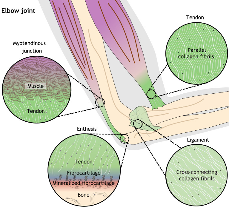
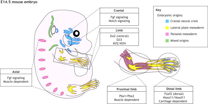
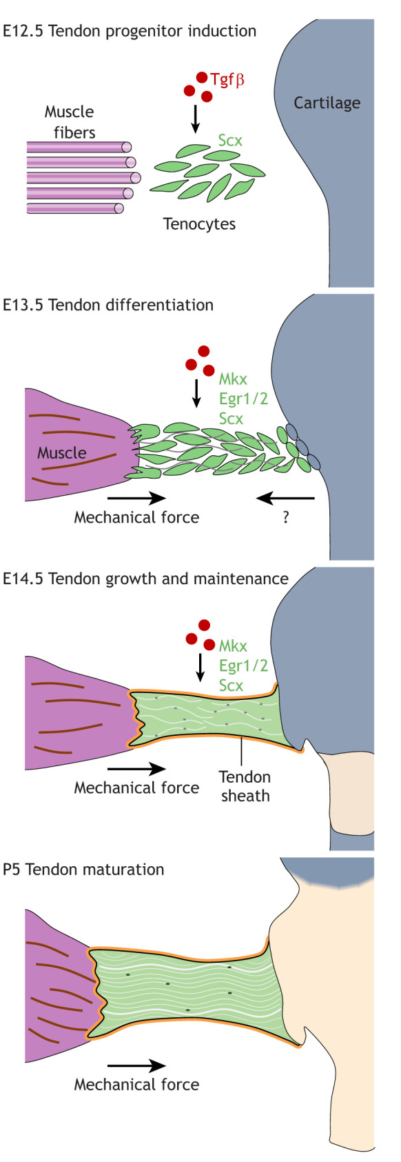
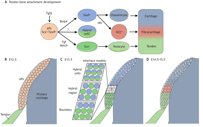
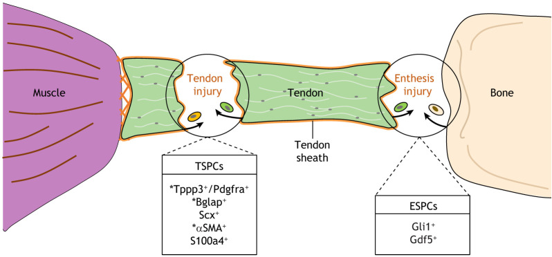
Tendons and ligaments are fibrous connective tissues vital to the transmission of force and stabilization of the musculoskeletal system. Arising in precise regions of the embryo, tendons and ligaments share many properties and little is known about the molecular differences that differentiate them. Recent studies have revealed heterogeneity and plasticity within tendon and ligament cells, raising questions regarding the developmental mechanisms regulating tendon and ligament identity. Here, we discuss recent findings that contribute to our understanding of the mechanisms that establish and maintain tendon progenitors and their differentiated progeny in the head, trunk and limb. We also review the extent to which these findings are specific to certain anatomical regions and model organisms, and indicate which findings similarly apply to ligaments. Finally, we address current research regarding the cellular lineages that contribute to tendon and ligament repair, and to what extent their regulation is conserved within tendon and ligament development.
Keywords: Enthesis; Fgf; Fibrous connective tissue; Ligament; Scx; Tendon; Tenocyte.
© 2021. Published by The Company of Biologists Ltd.
Conflict of interest statement
Competing interests The authors declare no competing or financial interests.
Figures
Similar articles
-
The development of zebrafish tendon and ligament progenitors.Development. 2014 May;141(10):2035-45. doi: 10.1242/dev.104067. Development. 2014. PMID: 24803652 Free PMC article.
-
TGFβ and FGF promote tendon progenitor fate and act downstream of muscle contraction to regulate tendon differentiation during chick limb development.Development. 2016 Oct 15;143(20):3839-3851. doi: 10.1242/dev.136242. Epub 2016 Sep 13. Development. 2016. PMID: 27624906
-
Divergent differentiation of skeletal progenitors into cartilage and tendon: lessons from the embryonic limb.ACS Chem Biol. 2014 Jan 17;9(1):72-9. doi: 10.1021/cb400713v. Epub 2013 Nov 20. ACS Chem Biol. 2014. PMID: 24228739 Review.
-
Tendon and ligament: development, repair and disease.Birth Defects Res C Embryo Today. 2005 Sep;75(3):226-36. doi: 10.1002/bdrc.20049. Birth Defects Res C Embryo Today. 2005. PMID: 16187327 Review.
-
Mechanism of muscle-tendon-bone complex development in the head.Anat Sci Int. 2020 Mar;95(2):165-173. doi: 10.1007/s12565-019-00523-0. Epub 2020 Jan 8. Anat Sci Int. 2020. PMID: 31916224 Review.
Cited by
-
New developments in the biology of fibroblast growth factors.WIREs Mech Dis. 2022 Jul;14(4):e1549. doi: 10.1002/wsbm.1549. Epub 2022 Feb 9. WIREs Mech Dis. 2022. PMID: 35142107 Free PMC article. Review.
-
Advanced Gene Therapy Strategies for the Repair of ACL Injuries.Int J Mol Sci. 2022 Nov 21;23(22):14467. doi: 10.3390/ijms232214467. Int J Mol Sci. 2022. PMID: 36430947 Free PMC article. Review.
-
Genetically engineered zebrafish as models of skeletal development and regeneration.Bone. 2023 Feb;167:116611. doi: 10.1016/j.bone.2022.116611. Epub 2022 Nov 14. Bone. 2023. PMID: 36395960 Free PMC article. Review.
-
Hemiepiphysiodesis Corrects Lower Extremity Coronal Plane Deformity in Children with Skeletal Dysplasia Irrespective of Intra-Articular Malalignment.J Pediatr Soc North Am. 2024 Jun 26;8:100068. doi: 10.1016/j.jposna.2024.100068. eCollection 2024 Aug. J Pediatr Soc North Am. 2024. PMID: 40433003 Free PMC article.
-
Transcriptome profiling of tendon fibroblasts at the onset of embryonic muscle contraction reveals novel force-responsive genes.Elife. 2025 Mar 27;14:e105802. doi: 10.7554/eLife.105802. Elife. 2025. PMID: 40145570 Free PMC article.
References
Publication types
MeSH terms
Grants and funding
LinkOut - more resources
Full Text Sources
Medical
