. 2021 Apr;51(2):100-113.
doi: 10.5051/jpis.2005120256.

Compromised extraction sockets: a new classification and prevalence involving both soft and hard tissue loss

Affiliations

Compromised extraction sockets: a new classification and prevalence involving both soft and hard tissue loss

Jung Ju Kim et al. J Periodontal Implant Sci. 2021 Apr.

Abstract

Purpose: Previous studies have solely focused on fresh extraction sockets, whereas in clinical settings, alveolar sockets are commonly associated with chronic inflammation. Because the extent of tissue destruction varies depending on the origin and the severity of inflammation, infected alveolar sockets may display various configurations of their remaining soft and hard tissues following tooth extraction. The aim of this study was to classify infected alveolar sockets and to provide the appropriate treatment approaches.

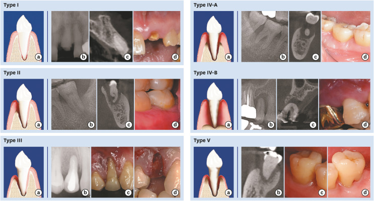
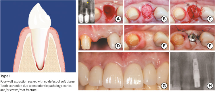
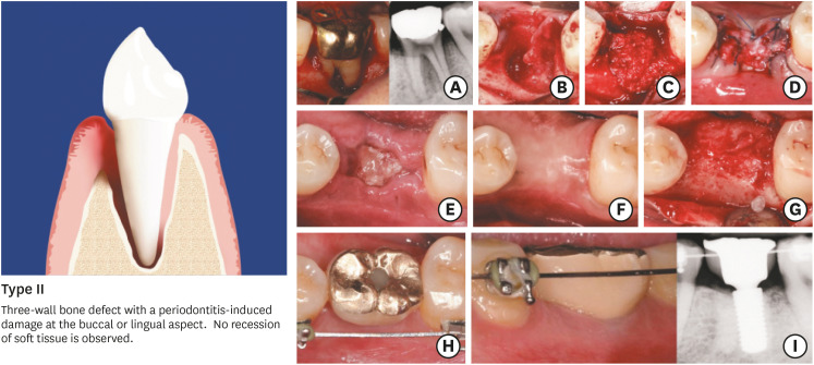
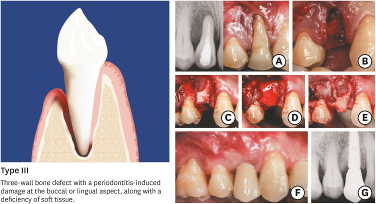
Methods: A proposed classification of extraction sockets with chronic inflammation was developed based upon the morphology of the bone defect and soft tissue at the time of tooth extraction. The prevalence of each type of the suggested classification was determined retrospectively in a cohort of patients who underwent, between 2011 and 2015, immediate bone grafting procedures (ridge preservation/augmentation) after tooth extractions at Seoul National University Dental Hospital.

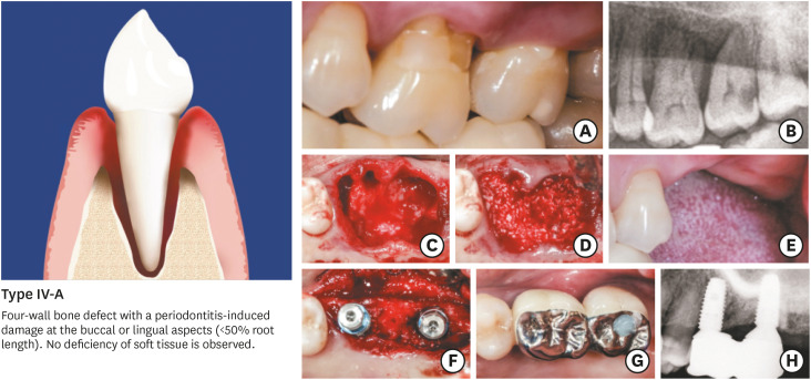
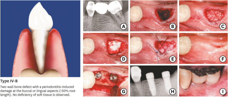
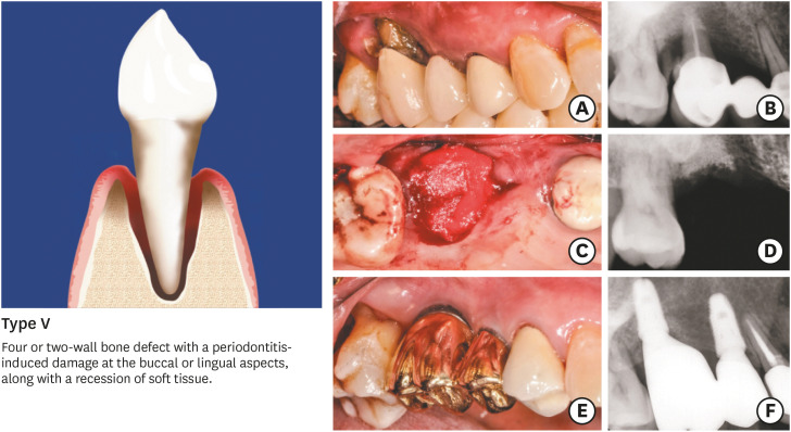
Results: The extraction sockets were classified into 5 types: type I, type II, type III, type IV (A & B), and type V. In this system, the severity of bone and soft tissue breakdown increases from type I to type V, while the reconstruction potential and treatment predictability decrease according to the same sequence of socket types. The retrospective screening of the included extraction sites revealed that most of the sockets assigned to ridge preservation displayed features of type IV (86.87%).

Conclusions: The present article classified different types of commonly observed infected sockets based on diverse levels of ridge destruction. Type IV sockets, featuring an advanced breakdown of alveolar bone, appear to be more frequent than the other socket types.

Keywords: Alveolar ridge augmentation; Classification; Periodontitis; Socket graft; Tooth socket.


Conflict of interest statement

No potential conflict of interest relevant to this article was reported.

Figures

Figure 1
Figure 1. Classification of compromised sockets and clinical and radiographic presentation of socket types.
Type I socket. A 4-wall socket without the resorption of the socket wall. Schematic drawing (a), radiographic periapical image (b), computed tomographic image (c) and clinical photo (d) (#21 tooth area). Type II socket. A 3-wall defect with resorption of either the buccal or palatal (or lingual) wall and no soft tissue recession. Schematic drawing (a), radiographic periapical image (b), computed tomographic image (c), and clinical photo (d) (#37 tooth area). Type III socket. A 3-wall defect with resorption of either the buccal or palatal (or lingual) wall and soft tissue recession. Schematic drawing (a), radiographic periapical image (b), clinical photo before tooth extraction (c), clinical photo after tooth extraction showing the intact palatal wall and soft tissue (d) (#14 tooth area). Type IV-A socket. Less than 50% of bone resorption on both buccal and palatal (or lingual) walls and soft tissue in the normal position. Schematic drawing (a), radiographic periapical image (b), computed tomographic image (c), and clinical photo (d) (#47 tooth area). Type IV-B socket. A 2-wall defect with more than 50% buccal and palatal (or lingual) bone loss and soft tissue in the normal position. Schematic drawing (a), radiographic periapical image (b), computed tomographic image (c) and clinical photo (d) (#15 tooth area). Type V socket. Bone resorption and gingival recession of soft tissue on both buccal and palatal (or lingual) walls. Schematic drawing (a), computed tomographic image (b), clinical photo of the buccal side (c), and clinical photo of the lingual side (d) (#47 tooth area).
Figure 2
Figure 2. Type I. (A) Infected extraction socket presenting features of type I: a 4-wall socket without the resorption of socket walls with an endodontic lesion. (B) Application of bone graft after debridement. (C) The socket entrance is sealed with a collagen matrix and secured with sutures. (D) Healing at postoperative week 2. (E) Ridge contour 4 months after ridge preservation. (F) Implant insertion. (G, H) Follow-up at 1 year after prosthesis delivery shows favorable aesthetic results and a stable radiographic level of peri-implant marginal bone.
Figure 3
Figure 3. Type II. (A) Advanced periodontitis resulting into a substantial defect of the buccal bone wall with endodontic involvement. (B) Damaged extraction socket in line with the presentation of type II. (C) Ridge preservation with deproteinized bovine bone minerals and native collagen membrane. (D) Sutures after socket grafting. (E) Healing at 2 weeks following surgery. (F, G) Ridge preservation allowed implant installation. (H, I) Follow-up at 3 years after prosthesis delivery shows favorable aesthetic results and a stable radiographic level of peri-implant marginal bone.
Figure 4
Figure 4. Type III. (A) Periodontitis-induced bone damage at the buccal wall, along with a recession on tooth #14. (B) A damaged extraction socket shows the presentation of type III marked by a soft tissue deficiency. (C) Socket grafting using deproteinized bovine bone minerals with 10% collagen. (D) Insertion of a native collagen membrane as a barrier device. (E) A connective tissue graft inserted onto the buccal mucosa. (F) Clinical and radiographic outcomes at 1 year after functional loading.
Figure 5
Figure 5. Type IV-A. (A, B) Clinical and radiographic features of type IV-A in tooth #26: buccal and palatal bone resorption (<50% of root length) with no defect of soft tissues, along with recession on tooth #14. (B) Extraction of teeth #26 and #27 followed by a meticulous debridement under copious saline irrigation. (D) Socket grafting using deproteinized bovine bone mineral with 10% collagen prior to coverage with a native collagen membrane. (E) Ridge healing 3 months following surgery. (F–H) Re-entry for implant insertion into a proper bone envelope, followed by prosthetic treatment and radiographic examination at 1 year following functional loading.
Figure 6
Figure 6. Type IV-B. (A) Advanced periodontitis on tooth #37 requiring tooth extraction. (B) The extraction socket displayed features of type IV-B (buccal and palatal bone resorption >50% of the root length, no soft tissue defect). (C) Socket grafting using deproteinized bovine bone mineral with 10% collagen. (D, E) Insertion of a native collagen membrane and sutures. (F) Healing at 3 months following surgery. (G, H) Implant installation. (I) Follow-up at 1 year after prosthesis delivery.
Figure 7
Figure 7. Type V. (A) Tooth #16 requiring extraction, with bone and soft tissue deficiency corresponding to type V. (B) Radiographic image at baseline. (C) Ridge preservation/augmentation using deproteinized bovine bone mineral with 10% collagen covered with a native collagen membrane. (D) Radiographic follow-up at 4 months after extraction. (E) Clinical situation at 1 year after implant loading. (F) Radiographic view 1 year after implant loading, documenting a stable level of peri-implant marginal bone.

References

    1. Pietrokovski J, Massler M. Alveolar ridge resorption following tooth extraction. J Prosthet Dent. 1967;17:21–27. - PubMed
    1. Schropp L, Wenzel A, Kostopoulos L, Karring T. Bone healing and soft tissue contour changes following single-tooth extraction: a clinical and radiographic 12-month prospective study. Int J Periodontics Restorative Dent. 2003;23:313–323. - PubMed
    1. Botticelli D, Berglundh T, Lindhe J. Hard-tissue alterations following immediate implant placement in extraction sites. J Clin Periodontol. 2004;31:820–828. - PubMed
    1. Tan WL, Wong TL, Wong MC, Lang NP. A systematic review of post-extractional alveolar hard and soft tissue dimensional changes in humans. Clin Oral Implants Res. 2012;23(Suppl 5):1–21. - PubMed
    1. Kim JH, Susin C, Min JH, Suh HY, Sang EJ, Ku Y, et al. Extraction sockets: erratic healing impeding factors. J Clin Periodontol. 2014;41:80–85. - PubMed

LinkOut - more resources