. 2021 Apr 29:10:e64457.
doi: 10.7554/eLife.64457.

The causal role of auditory cortex in auditory working memory

Affiliations

The causal role of auditory cortex in auditory working memory

Liping Yu et al. Elife. .

Abstract

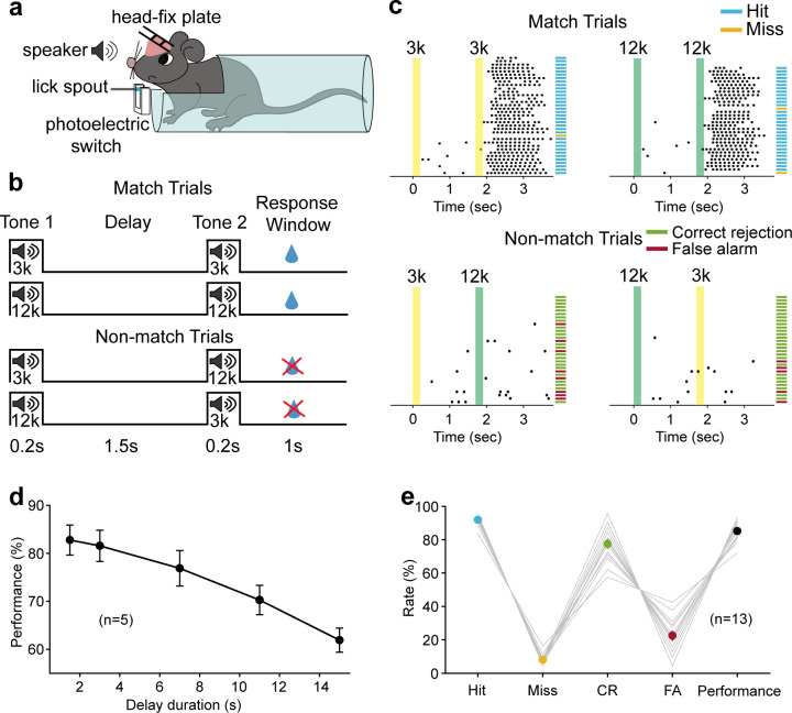
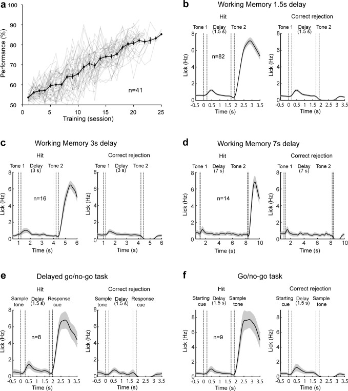
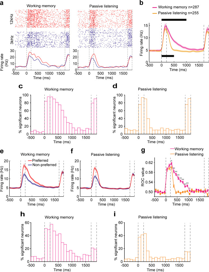
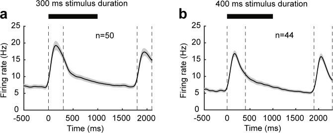
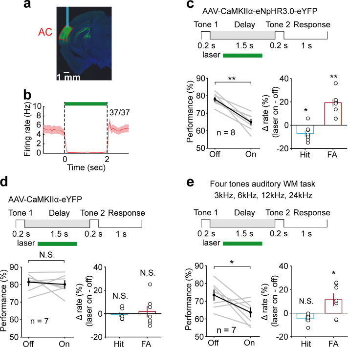
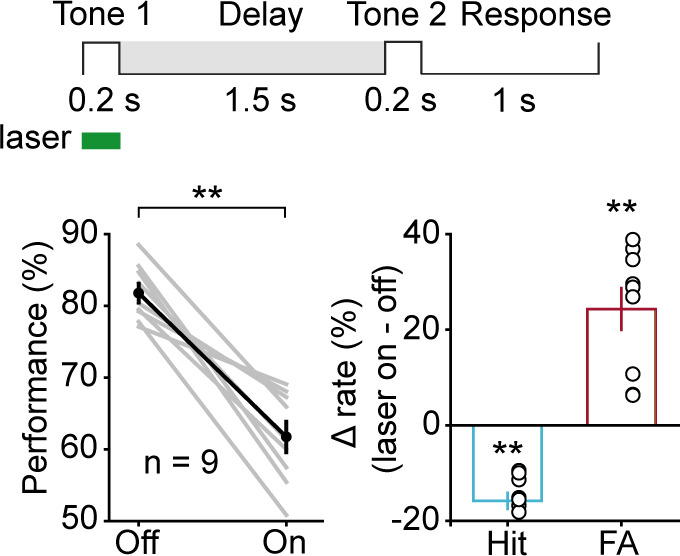
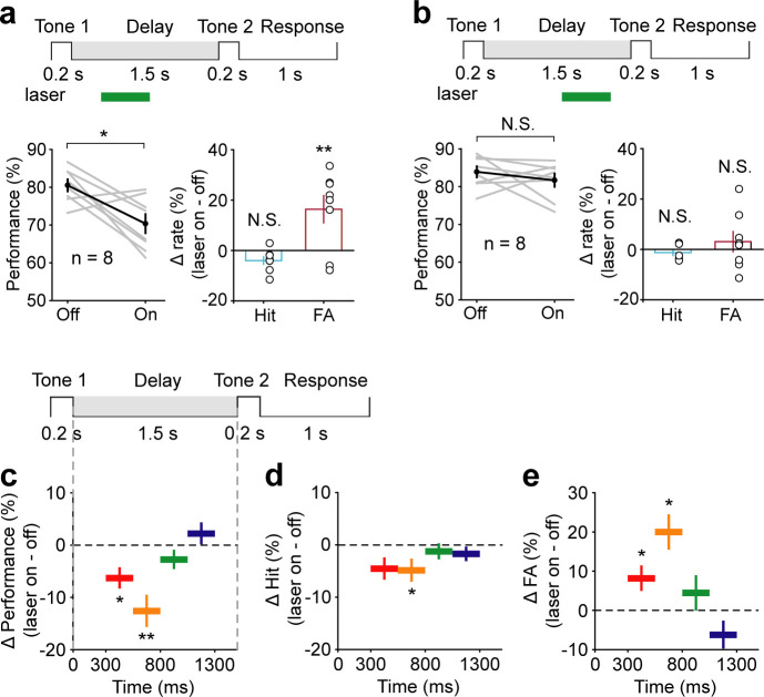
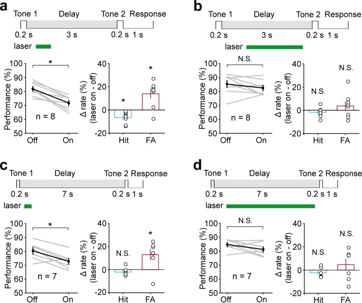
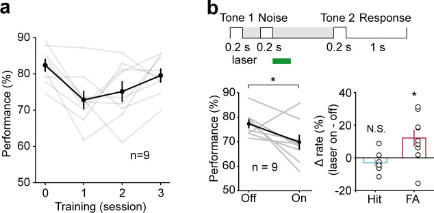
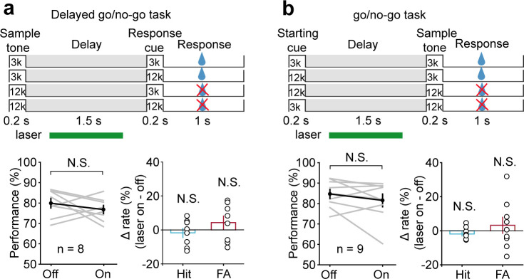
Working memory (WM), the ability to actively hold information in memory over a delay period of seconds, is a fundamental constituent of cognition. Delay-period activity in sensory cortices has been observed in WM tasks, but whether and when the activity plays a functional role for memory maintenance remains unclear. Here, we investigated the causal role of auditory cortex (AC) for memory maintenance in mice performing an auditory WM task. Electrophysiological recordings revealed that AC neurons were active not only during the presentation of the auditory stimulus but also early in the delay period. Furthermore, optogenetic suppression of neural activity in AC during the stimulus epoch and early delay period impaired WM performance, whereas suppression later in the delay period did not. Thus, AC is essential for information encoding and maintenance in auditory WM task, especially during the early delay period.

Keywords: auditory cortex; mice; mouse; neuroscience; working memory.

Plain language summary

Working memory is the ability to hold information in your head for a few seconds while making decisions, planning or applying logical reasoning to problem solving. It is a fundamental component of cognition, and yet it remains unclear where working memory is stored in the brain. The prefrontal cortex – the front lobe of the brain – is likely the main hub of working memory, since it is responsible for executive functions, such as decision making and planning. This idea is supported by experiments showing sustained brain activity in the prefrontal cortex during working memory tasks. Lesions in that part of the brain also lead to profound deficits in working memory. However, there is increasing evidence that other parts of the brain which process sensory information also participate in retaining working memory. The auditory cortex, which processes sound, is one such candidate. To find out whether the auditory cortex has a role to play in working memory, Yu, Hu, Shi et al. trained mice to lick a water spout after hearing the same sound twice in a row, 1.5 seconds apart, and then measured the activities of the mice’s neurons. This showed that neurons in the auditory cortex were active not only when the mice were presented with sound cues, but also for a short time during the delay period between sounds. Yu, Hu, Shi et al. then manipulated this neurons to inactivate them for a fraction of a second after the first sound, which resulted in the animals’ working memory was impaired. However, suppressing the activity of the auditory cortex cells in the later stages of the sound delay period had no effect on working memory. These results indicate that although the auditory cortex may not be involved in storing information for the entire working memory process, it is crucial for encoding of auditory information. In summary, this work uncovers how neurons in the auditory cortex underlie working memory. Further research focusing on these neurons could explain how working memory deteriorates with age, or why it is impaired in people with learning difficulties.


Conflict of interest statement

LY, JH, CS, LZ, MT, JZ, JX No competing interests declared

Figures

Figure 1.
Figure 1.. Auditory working memory task in head-fixed mice and behavioral performance.
(a) Diagram of the experimental setup. (b) Schematic for task design. For each trial, an auditory stimulus (3 kHz, or 12 kHz, 0.2 s) was presented as the sample, followed by a delay period of 1.5 s and a testing auditory stimulus (0.2 s), either matched or nonmatched to the sample. Mice were rewarded with water if they licked within a response window in the match trials. (c) Licking behavior in an example session and definition of the trial type. Colored areas correspond to the two auditory stimulus delivery periods, as indicated above. Each tick indicates one lick. Short horizontal lines indicate the trial types (blue: hit; orange: miss; green: correct rejection [CR]; magenta: false alarm [FA]). (d) The performance with varying delay duration (n = 5 mice). Mean ± s.e.m. (e) Mean hit, miss, CR, FA rates, and the performance of all mice (n = 13 mice) during neural recording sessions. Gray lines: individual mice; black: mean ± s.e.m. Hit + miss = 100%; CR + FA = 100%. See Figure 1—source data 1 for more details.
Figure 1—figure supplement 1.
Figure 1—figure supplement 1.. The learning process of the auditory working memory (WM) task and the licking behavior of well-trained mice.
(a) Performances of all mice while learning the auditory WM task of 1.5 s delay. (b–f) Averaged licking rate in hit (left) and correct rejection (right) trials of mice well trained for the WM task of 1.5 s delay (b), WM task of 3 s delay (c), WM task of 7 s delay (d), delayed go/no-go task (e), and go/no-go task (f).
Figure 2.
Figure 2.. Neural correlates of the auditory cortex activity in the auditory working memory (WM) task.
(a) Raster (top) and peri-stimulus time histograms (bottom) of an example neuron recorded during the WM behavior (left) and passive listening (right). Trials were sorted by auditory samples. The sample stimulus and test stimulus times are bounded by the vertical dotted lines. (b) Averaged population firing rates for neurons recorded during WM behavior (n = 287) and passive listening (n = 255). Shadows: s.e.m.; the black block on the top indicates the successive 100 ms bins with firing rate significantly different from baseline (500 ms before the beginning of sample) for neurons recorded during WM behavior. p<0.05, Wilcoxon rank-sum test. The black block below indicates significant bins for neurons recorded during passive listening. (c, d) Percentage of neurons with a significant difference in firing rate compared with baseline at different time points during WM behavior (c) and passive listening (d). (e, f) Averaged population firing rates for neurons recorded during WM behavior (e) and passive listening (f). Trials in which the sample stimulus was the preferred or nonpreferred, which varied for each neuron, are shown separately. (g) The average of receiver operating characteristic (ROC) values across populations, calculated in each 100 ms window, is plotted as a function of time for neurons recorded during WM behavior (magenta) and passive listening (orange). *p<0.05, **p<0.001, Wilcoxon rank-sum test. (h, i) Incidence of neurons with significant ROC values for each 100 ms epoch in WM behavior (h) and passive listening (i). p<0.05, permutation test.
Figure 2—figure supplement 1.
Figure 2—figure supplement 1.. Neural correlates of the auditory cortex activity in the auditory working memory (WM) task with varied stimulus duration.
(a, b) Averaged population firing rates for neurons recorded during auditory WM behavior with the stimulus duration of 300 ms (a) and 400 ms (b). The black block on the top indicates the successive 100 ms bins with firing rate significantly different from baseline (500 ms before the beginning of sample), p<0.05, Wilcoxon rank-sum test.
Figure 3.
Figure 3.. Suppression of delay-period activity in auditory cortex (AC) by optogenetic inhibition of pyramidal neurons impaired auditory working memory (WM) performance.
(a) Histology image showing the expression of AAV-CaMKIIα-eNpHR3.0-eYFP in AC. (b) Activity suppression efficiency revealed by optetrode recording in vivo. (c) Suppressing AC activity during the delay period of WM task decreased performance, with a substantial increase in false alarm (FA) rate and a small decrease in hit rate. Top: schematic of optogenetic stimulation during the delay period of WM task. The green rectangle indicates the period of inactivation. For the bottom panel, gray lines indicate individual mice; black indicates mean ± s.e.m. Circles indicate individual mice. *p<0.05, **p<0.001, t-test. (d) As in (c) with control virus injection. The photostimulation of AC with control virus injection during the delay period did not affect the behavior. N.S.: not significant. (e) Suppressing AC delay-period activity decreased the performance in the four tones auditory WM task with a decrease in FA rate and no change in hit rate. See Figure 3—source data 1 for more details.
Figure 3—figure supplement 1.
Figure 3—figure supplement 1.. Optogenetic suppression of auditory cortex during the stimulus epoch dramatically reduced the animals’ ability to perform the working memory task.
Figure 4.
Figure 4.. Temporal specificity of the effect of auditory cortex (AC) suppression.
(a) AC suppression during the delay period of 300–800 ms decreased the working memory (WM) behavioral performance, with an increase in false alarm (FA) rate and no change in hit rate. (b) AC suppression during the delay period of 800–1300 ms did not affect behavioral performance. (c–e) The WM behavioral change caused by AC suppression during the delay period of 300–550 ms (red; n = 8 mice), 550–800 ms (yellow; n = 8 mice), 800–1050 ms (green; n = 7 mice), and 1050–1300 ms (blue; n = 8 mice). The task structure is shown at the top. For the bottom panels, the horizontal extent of the colored bars indicates the period of inactivation. The vertical position indicates the average change in performance (c), hit rate (d), and FA rate (e) across mice induced by the corresponding period of AC suppression. Error bars show s.e.m. across mice. See Figure 4—source data 1 for more details.
Figure 5.
Figure 5.. Performance was impaired following optogenetic suppression of auditory cortex (AC) activity during the early delay period, with the delay duration of 3 s and 7 s.
(a, b) In working memory (WM) task with the delay duration of 3 s, AC suppression during the early delay period (300–800 ms) (a) but not later (800–2700 ms) (b) decreased the behavioral performance. (c, d) In WM task with the delay duration of 7 s, AC suppression during the delay period of 300–800 ms (c) but not 800–6700 ms (d) decreased the behavioral performance. See Figure 5—source data 1 for more details.
Figure 6.
Figure 6.. Active memory maintenance in auditory working memory (WM) task by the auditory cortex (AC) delay-period activity.
(a) Learning curve for the performance in the WM task with noise distractor (presented during 300–500 ms of the delay period). Note the drop of performance after inserting the noise distractor in the delay period on the first day. After 2 days of training, the performance data from the third day of the WM task with noise distractor was no worse than that in the simple WM task (the zeroth day). (b) Optogenetic suppression of AC during the early delay period after the distractor (500–800 ms) resulted in impairment in task performance.
Figure 7.
Figure 7.. Suppressing auditory cortex (AC) activity did not affect behavioral performance in the delayed go/no-go auditory discrimination task and go/no-go auditory discrimination task.
(a) Paradigm and behavioral performance for the delayed go/no-go auditory discrimination task experiments with suppressed AC activity. (b) As in (a) for the go/no-go auditory discrimination task. See Figure 6—source data 1 for more details.

Similar articles

Cited by

References

    1. Baddeley A. Working memory. Science. 1992;255:556–559. doi: 10.1126/science.1736359. - DOI - PubMed
    1. Baddeley A. Working memory: theories, models, and controversies. Annual Review of Psychology. 2012;63:1–29. doi: 10.1146/annurev-psych-120710-100422. - DOI - PubMed
    1. Bettencourt KC, Xu Y. Decoding the content of visual short-term memory under distraction in occipital and parietal Areas. Nature Neuroscience. 2016;19:150–157. doi: 10.1038/nn.4174. - DOI - PMC - PubMed
    1. Bigelow J, Rossi B, Poremba A. Neural correlates of short-term memory in primate auditory cortex. Frontiers in Neuroscience. 2014;8:250. doi: 10.3389/fnins.2014.00250. - DOI - PMC - PubMed
    1. Chafee MV, Goldman-Rakic PS. Matching patterns of activity in primate prefrontal area 8a and parietal area 7ip neurons during a spatial working memory task. Journal of Neurophysiology. 1998;79:2919–2940. doi: 10.1152/jn.1998.79.6.2919. - DOI - PubMed

Publication types

LinkOut - more resources