

A novel approach to anterior segment imaging with smartphones in the COVID-19 era
- PMID: 33913872
- PMCID: PMC8186572
- DOI: 10.4103/ijo.IJO_3707_20
A novel approach to anterior segment imaging with smartphones in the COVID-19 era
Abstract
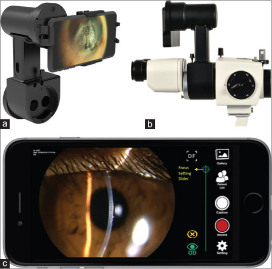
Purpose: To report a novel, telemedicine-friendly, smartphone-based, wireless anterior segment device with instant photo-documentation ability in the COVID-19 era.
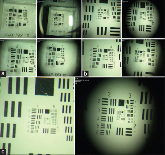
Methods: Anterior Imaging Module (AIM) was constructed based on a 50/50 beam splitter design, to match the magnification drum optics of slit-lamps with a three-step or higher level of magnification. The design fills the smartphone sensor fully at the lowest magnification and matches the fixed focus of the slit-lamp. It comes with a smartphone for instant photo-documentation, an in-built software application for data-management and secure HIPAA compliant cloud storage, and a Bluetooth trigger for a one-tap image capture. The construction of the device is explained, and the optical resolution measured using U.S. air-force resolution test. AIM's performance was characterized with traceability to internationally relevant performance standards for digital slit-lamps after image quality assessment through a pilot study.
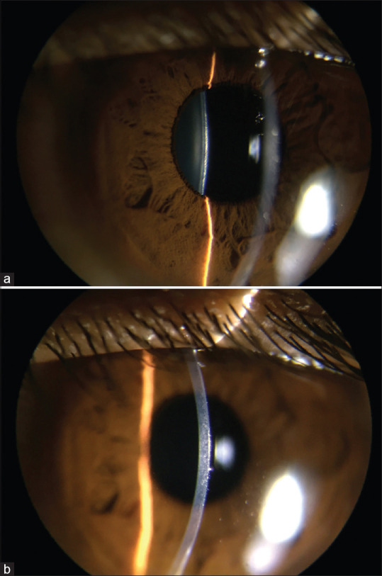
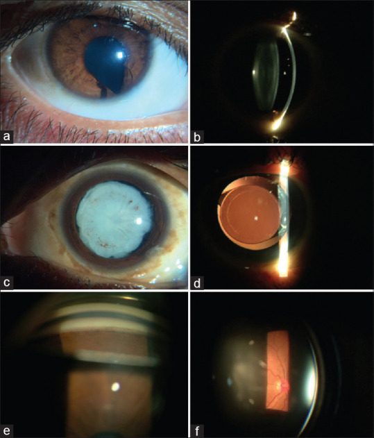
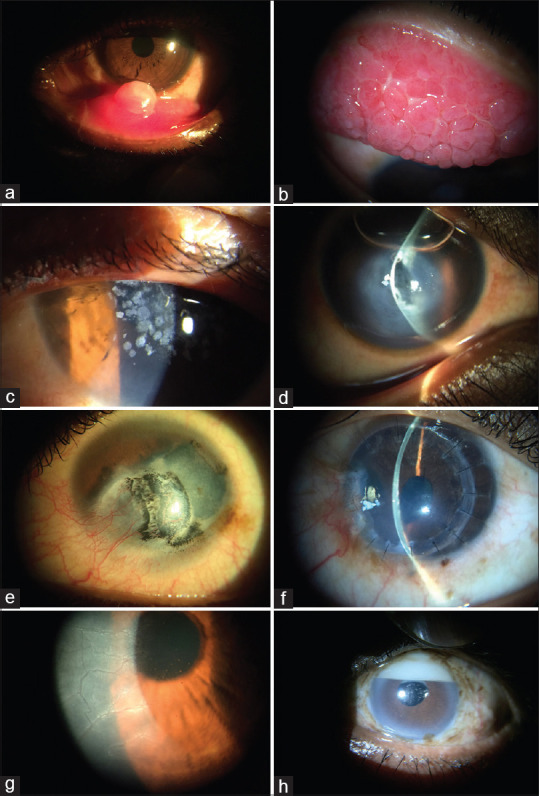
Results: Clinically useful anterior segment images were obtained with both diffuse and slit illumination at different magnification settings with the highest magnification (40X) resolution of 359 lines per cm and the lowest magnification (16X) resolution of 113 lines per cm.
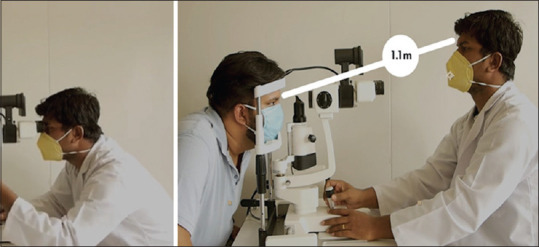
Conclusion: AIM is a novel, wireless, telemedicine-enabled design that digitizes existing, analog slit lamps with at least three-step magnification. The settings ensure the focus is determined purely by the position of the slit-lamp. Hence, the image viewed and captured on the smartphone is exactly what the clinician sees through the eyepiece. This helps in maintaining distance from the patient in the ongoing COVID-19 pandemic, as well.
Keywords: Anterior segment imaging; COVID-19; conjunctiva; cornea; iris; lens; sclera; slit-lamp; smartphone; telemedicine; uniform illumination.
Conflict of interest statement
None
Figures

Comment in
-
Commentary: Smartphone-based, wireless, slit-lamp imaging with small footprint - making photodocumentation easy.Indian J Ophthalmol. 2021 May;69(5):1262-1263. doi: 10.4103/ijo.IJO_796_21. Indian J Ophthalmol. 2021. PMID: 33913873 Free PMC article. No abstract available.
References
-
- Oliphant H, Kennedy A, Comyn O, Spalton DJ, Nanavaty MA. Commercial slit lamp anterior segment photography versus digital compact camera mounted on a standard slit lamp with an adapter. Curr Eye Res. 2018;43:1290–4. - PubMed
-
- Ye Y, Jiang H, Zhang H, Karp CL, Zhong J, Tao A, et al. Resolution of slit-lamp microscopy photography using various cameras. Eye Contact Lens. 2013;39:205–13. - PubMed
MeSH terms
LinkOut - more resources
Full Text Sources
Other Literature Sources
Medical
Miscellaneous
