. 2021 May;69(5):1275-1282.
doi: 10.4103/ijo.IJO_403_21.

Retinal manifestations in patients following COVID-19 infection: A consecutive case series

Affiliations

Retinal manifestations in patients following COVID-19 infection: A consecutive case series

Mallika Goyal et al. Indian J Ophthalmol. 2021 May.

Abstract

Purpose: To describe retinal manifestations seen in patients associated with COVID-19 infection at a multi-specialty tertiary care hospital in Southern India.

Methods: In this retrospective chart review, all consecutive cases presenting to the Retina-Uveitis service from May 2020 to January 2021 with retinal manifestations associated with COVID-19 infection or its sequelae or as a result of treatment given for COVID-19 were included.

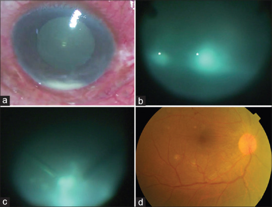
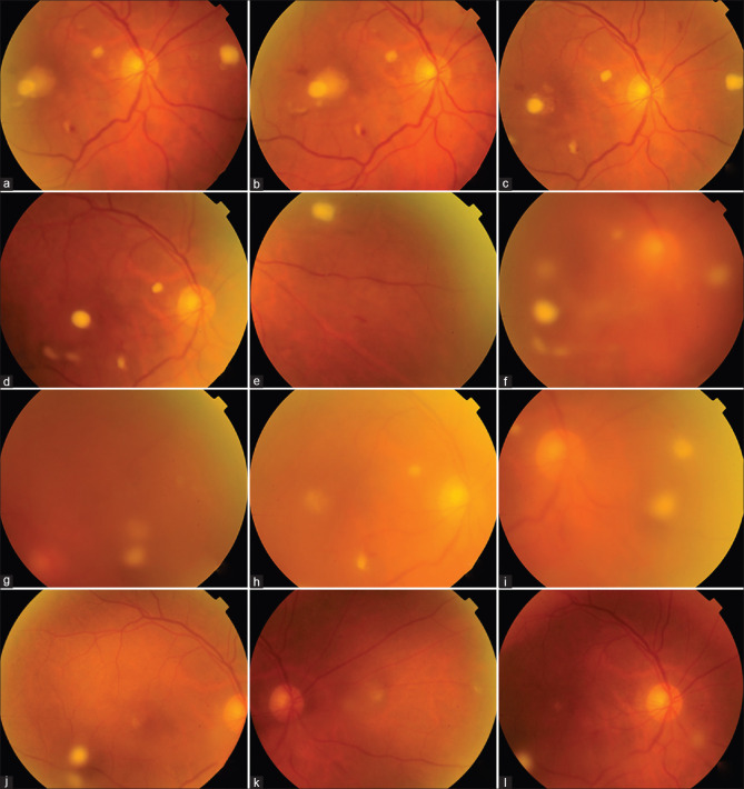
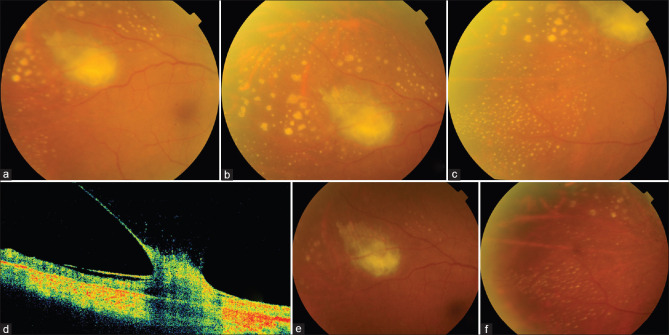
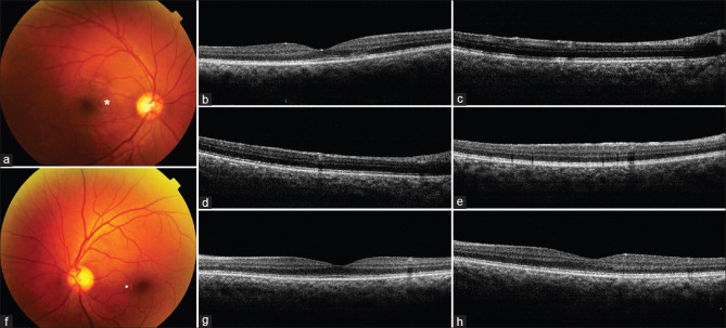
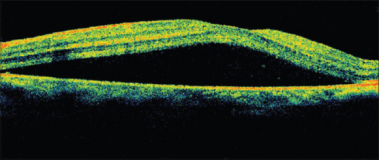
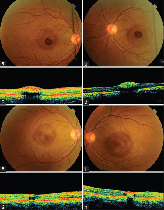
Results: : Of the 7 patients, 3 were female, and 4 were male. Four patients had onset of symptoms during the active phase of COVID-19 infection. Four had bilateral and three had unilateral involvement. The manifestations ranged from mild to vision threatening. Vision threatening manifestations included infections: endogenous endophthalmitis, candida retinitis and tubercular choroidal abscess and bilateral pre-foveal hemorrhages. Milder manifestations included paracentral acute middle maculopathy, central serous chorio-retinopathy and voriconazole induced visual symptoms. Final visual acuity was 6/36 or better in the four severe cases and 6/9 or better in the mild cases.

Conclusion: This study highlights the retinal manifestations associated with COVID-19 infection and its sequelae. As these patients presented with an association with COVID-19 (either during or after recovery), ophthalmologists should be vigilant and screen for such entities in case of complaints of visual symptoms or in the presence of systemic sepsis. The outcomes can be good with prompt and aggressive management.

Keywords: COVID 19; COVID-associated mycoses; endophthalmitis; ocular manifestations; paracentral acute middle maculopathy; retinal manifestations.


Conflict of interest statement

None

Figures

Figure 1
Figure 1
(a) Intraoperative photograph of the right eye shows severe ciliary congestion and hypopyon. (b) Intra-operative photograph during pars plana vitrectomy shows dense and dry vitreous lesions (asterisk) (c) Snowball like opacities noted in the inferior periphery (d) Same eye shows compete resolution with clear media and normal fundus at 6 weeks
Figure 2
Figure 2
(a-k) Creamy-white candida retinitis lesions at presentation (a), macular involvement (b) Two weeks after voriconazole (c) Six weeks later: increasing activity (d and e) 10 days after intravitreal injections new vitreous exudates are noted (f); older retinitits lesions show some resolution (g)The lesions and vitreous exudates persist with some resolution after 4 weeks of injections. 3 months review: complete resolution of vitreous inflammation and retinitis, (j-l) with scarring noted at the site of the original lesions
Figure 3
Figure 3
(a) Large choroidal abscess with active appearing center and resolving activity at edges supero-temporal to the right macula. (b and c) Multiple miliary lesions around the choroidal abscess in temporal and inferotemporal fundus. Lesions are larger and irregular closer to the abscess, becoming smaller, punctate with smoother edges further from it. (d) OCT showing epiretinal membrane and vitreous traction over the choroidal abscess. (e and f) Significant resolution of choroidal abscess and miliary lesions 6 weeks after initiating anti-tubercular therapy
Figure 4
Figure 4
(a) Triangular greyish-white lesion (asterisk) of AMN (b) Corresponding OCT shows disruption of external retinal layers (c and d) Focal areas of hyper-reflectivity with deeper shadowing of PAMM. (e) OCT right eye: diffuse hyper-reflectivity of entire inner retinal surface (f) Small greyish-white lesions in superficial retina nasal, inferonasal and temporal to the foveal center in left eye. (g) Corresponding OCT shows hyper-reflective lesion with underlying shadowing (h) OCT left eye showing diffuse hyper-reflectivity of the inner retinal surface
Figure 5
Figure 5
OCT left eye of a young female treated with oral steroids for COVID-19 showing serous macular detachment
Figure 6
Figure 6
Bilateral pre-foveal hemorrhage of the right (a) and left eye (b) in a young lady with post COVID-19 sepsis. OCT of the right eye (c) and left eye (d) showing pre-foveal location of the hemorrhage with underlying shadowing. One month later, there is almost complete resolution of the pre-foveal heme in the RE (e) and LE (f) Corresponding optical coherence tomography scans show residual paracentral pre-foveal heme in the RE (g) and intra-retinal heme in the LE (h)

Comment in

References

    1. Bertoli F, Veritti D, Danese C, Samassa F, Sarao V, Rassu N, et al. Ocular findings in COVID-19 patients:A review of direct manifestations and indirect effects on the eye. J Ophthalmol. 2020;2020:4827304. doi:10.1155/2020/4827304. - PMC - PubMed
    1. Douglas KA, Douglas VP, Moschos MM. Ocular manifestations of COVID-19 (SARS-CoV-2):A critical review of current literature. In Vivo. 2020;34(3 Suppl):1619–28. - PMC - PubMed
    1. Lani-Louzada R, Ramos CdVF, Cordeiro RM, Sadun AA. Retinal changes in COVID-19 hospitalized cases. PLoS One. 2020;15:e0243346. - PMC - PubMed
    1. Song G, Liang G, Liu W. Fungal co-infections associated with global COVID-19 pandemic:A clinical and diagnostic perspective from China. Mycopathologia. 2020;7:1–8. - PMC - PubMed
    1. Lei Y, Song Y, Shu Y, Zhao Y, Huo X, Wang H, et al. Fungal antigenemia in patients with severe Coronavirus disease 2019 (COVID-19):The facts and challenges. J Microbiol Immunol Infect. 2020;53:657–9. - PMC - PubMed