Review
. 2021 Apr 29;12(1):57.
doi: 10.1186/s13244-021-00997-5.

Image-guided percutaneous ablation for the treatment of lung malignancies: current state of the art

Affiliations
Review

Image-guided percutaneous ablation for the treatment of lung malignancies: current state of the art

Alfredo Páez-Carpio et al. Insights Imaging. .

Abstract

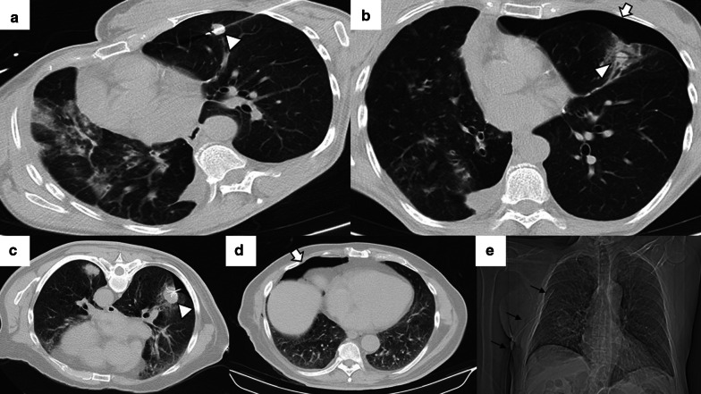
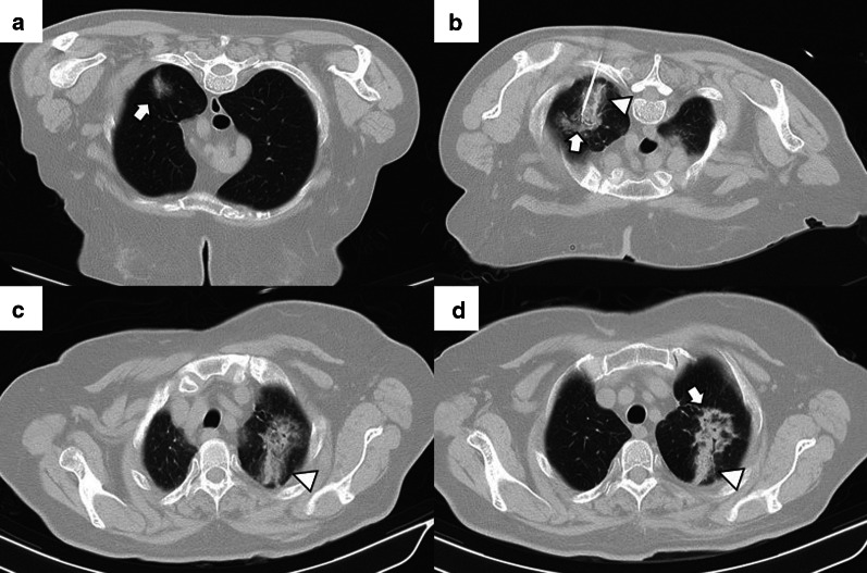
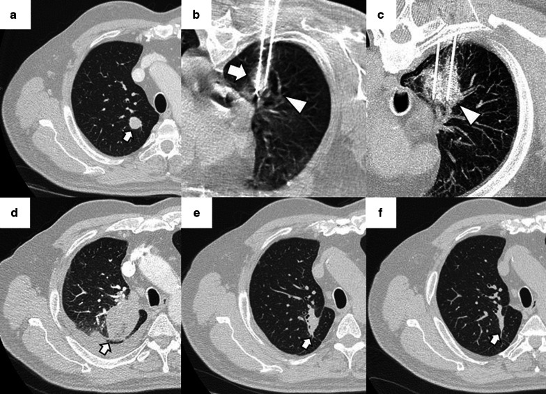
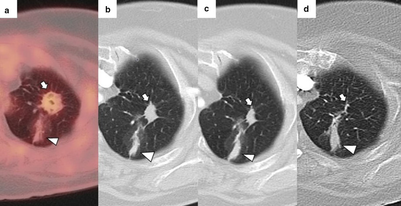
Image-guided percutaneous lung ablation has proven to be a valid treatment alternative in patients with early-stage non-small cell lung carcinoma or oligometastatic lung disease. Available ablative modalities include radiofrequency ablation, microwave ablation, and cryoablation. Currently, there are no sufficiently representative studies to determine significant differences between the results of these techniques. However, a common feature among them is their excellent tolerance with very few complications. For optimal treatment, radiologists must carefully select the patients to be treated, perform a refined ablative technique, and have a detailed knowledge of the radiological features following lung ablation. Although no randomized studies comparing image-guided percutaneous lung ablation with surgery or stereotactic radiation therapy are available, the current literature demonstrates equivalent survival rates. This review will discuss image-guided percutaneous lung ablation features, including available modalities, approved indications, possible complications, published results, and future applications.

Keywords: Metastatic lung disease; Non-small cell lung carcinoma; Percutaneous thermal ablation.


Conflict of interest statement

The authors declare that they have no competing interests.

Figures

Fig. 1
Fig. 1
Percutaneous access marking for CTF-guided lung ablation. a Preprocedural CT of a patient scheduled for treatment by image-guided percutaneous lung ablation of a single lung colon cancer metastasis located in the left upper lobe (arrow). Note that the CT scan was performed in the position chosen to perform the procedure (prone). b Marking of the lesion with a radiopaque grid (triangle). The scanner parameters were modified to decrease the exposure to ionizing radiation of the operator and the patient. Hence the decreased image quality. c Example of the marking procedure. The vertical axis is marked with the radio-opaque marker, while the horizontal axis is provided by the CT laser marking (acknowledgment to our resident, Dr. Tomás Fernández, for volunteering for the figure). d CTF-guided percutaneous access to the lesion. Note that the access coincides with the point marked in (b) (triangle)
Fig. 2
Fig. 2
Correct positioning of ablation probes in the target lesion, with schematic illustrations of each ablative alternative. a CT-guided percutaneous RFA treatment with a straight needle of a metastatic colon cancer lesion in the lower right lobe. Note the probe's correct positioning in the center of the tumor (arrow). b CT-guided percutaneous RFA treatment with an expandable needle of a colon cancer metastasis. The probe’s electrodes include the entire lesion (triangles). c CT-guided percutaneous MWA treatment with a single straight needle of a renal cancer metastatic lesion in the lower left lobe. As in (a), the probe passes through the center of the tumor (arrow). d CT-guided percutaneous CA of a stage I NSCLC in the lower right lobe with two straight ablation probes. If two or more ablation probes are used, they should be placed at the tumor's edges
Fig. 3
Fig. 3
Correct positioning of the ablation probe while treating a subpleural tumor. RFA of a subpleural nodular lesion in the lower right lobe, performed with a straight needle. Note that the needle makes a tangential route to the lesion (white arrow), evading the seemingly easier direct approach (triangle). This approach is necessary to allow a better anchorage in the lung parenchyma, thus avoiding the needle's non-voluntary displacement
Fig. 4
Fig. 4
Lung parenchyma retraction while performing an image-guided percutaneous lung ablation. a, b RFA treatment with a straight needle of a metastatic lesion of a colon adenocarcinoma located in the left upper lobe, very close to the pulmonary hilum (white arrow). c After treatment, the treated area presents an image with a consolidative center and a ground-glass peripheral halo, suggesting a complete treatment (white arrow). A discrete retraction of the pulmonary parenchyma is evident, which, added to the presence of a mild pneumothorax (white triangle), mobilizes the hilar structures. d Control of the treated lesion after 12 months of RFA, no signs of recurrence are evident
Fig. 5
Fig. 5
Mild pneumothorax after image-guided percutaneous lung ablation. a, b CT-guided MWA in a single lung metastasis from colon cancer in the upper left lobe. Note the correct probe position in the center of the lesion (triangle). In the CT performed immediately after the procedure, the patient presented a mild pneumothorax (arrow). The pneumothorax was resolved spontaneously without the need for a chest tube. Note the peripheral ground-glass halo with a central consolidation (triangle in b), which indicates a correct treatment. ce CT-guided RFA lung ablation in a lung metastasis from a basal cell skin carcinoma. The expandable probe correctly englobes the lesion (triangle in c). In this patient, a pneumothorax was also observed immediately after the ablation. (arrow in d). Although pneumothorax is similar to in A-B, the patient's clinical situation required a chest tube (black arrows in e)
Fig. 6
Fig. 6
Mild hemothorax after image-guided percutaneous lung ablation. a CT-guided percutaneous RFA of a stage 1 NSCLC located in the lower left lobe. Note the expanding tube encompassing the entire lesion. b CT scan immediately after the procedure where we observe a mild hyperdense pleural effusion, consistent with a mild hemothorax. c CT scan performed 24 h after the procedure shows a slight increase in the hyperdense pleural effusion and a discrete hyperdense level within the pleural effusion, confirming the hemothorax (arrow). However, after 48 h of clinical and radiological stability, the patient was discharged
Fig. 7
Fig. 7
Mild lung hemorrhage in the path of the ablation probe. a, b CT-guided percutaneous RFA with an expandable probe of a stage 1 NSCLC (arrow in a). The expandable probe completely covers the tumor in b (arrow), and the formation of a lung hemorrhage is visible in the probe’s path (triangle). c In the CT scan performed immediately after the procedure, we can observe a mild lung hemorrhage related to the probe’s path (triangle). d The lung hemorrhage remains stable in the CT 24 h after the procedure, and the patient remains asymptomatic. Note the presence of a ground-glass halo englobing a consolidative center, a suggestive sign of a correct ablative treatment (arrow in d)
Fig. 8
Fig. 8
Schematic illustration demonstrating the ground-glass halo's correct disposition surrounding the treated area. a Proper disposition of the ground-glass halo after ablation lung treatment. The halo must completely surround the tumor and be greater than 5 mm, preferably between 8 and 10 mm. bd Different forms of incomplete treatment after percutaneous pulmonary ablation as demonstrated by the disposition of the ground-glass halo
Fig. 9
Fig. 9
Radiological findings indicative of successful treatment immediately following image-guided percutaneous lung ablation. a CT-guided MWA with a single straight probe of a stage 1 NSCLC located in the lower right lobe. b We observed a consolidative center (black arrow) surrounded by a peripheral ground-glass halo (triangle) in the area treated in the CT scan performed immediately after completion of the procedure. The combination of these findings is indicative of successful treatment. c Follow-up CT one month after treatment. Note the increase in the size of the treated area compared to A. However, there are no irregular nodular areas nor other signs of tumor persistence. d CT 6 months after treatment The treated area has decreased in size compared to C, suggesting a successful treatment
Fig. 10
Fig. 10
Tumor persistence after treatment with image-guided percutaneous lung ablation. a Non-enhanced CT showing a single metastatic tumor in the upper right lobe in a patient with rectal carcinoma. b CT-guided RFA treatment using a straight probe, passing through the center of the tumor. c A non-enhanced CT was performed 24 h after treatment, showing the treated lesion (thick white arrow) surrounded by a ground-glass halo (triangle). However, the treated tumor's frontmost portion does not present a ground-glass halo (thin black arrow), which may correspond to incomplete treatment. d Contrast-enhanced CT scan performed three months after treatment shows a considerable increase in the treated lesion, which now presents nodular and irregular borders, consistent with tumor persistence
Fig. 11
Fig. 11
"Iceball" formation while performing an image-guided percutaneous lung CA. a CT of a patient with oligometastatic disease due to a small cell renal carcinoma with a lesion near the descending thoracic aorta (arrow). b CT-guided CA of this lesion, given the lesion's small size, it was possible to use a single straight probe placed in the center of the lesion (arrow). Note the hypodense halo surrounding the lesion, consistent with the “ice ball” formed during treatment (triangle). c Non-contrast CT scan one month after treatment. The treated area shows a larger size than the original lesion (arrow). However, it is a rounded area of similar size to the image immediately after treatment (not available). d Non-contrast CT scan six months after treatment. A lung scar is now observed in the treated area, consistent with effective treatment (arrow)
Fig. 12
Fig. 12
"Iceball" formation and further evolution to an area of lung hemorrhage in an image-guided percutaneous pulmonary cryoablation. a CT scan performed before ablative treatment, demonstrating a single metastatic lesion from a colon carcinoma in the upper right lobe (arrow). b, c CT-guided CA performed with two straight probes. Note the position of the probes, parallel and at the edges of the lesion. In (b) the ice ball (triangle) is observed surrounding the tumor (arrow). This ice ball was subsequently replaced by a ground-glass halo in (c) (triangle). d Contrast-enhanced CT scan performed one month after treatment, with a treated area more extensive than the original lesion (arrow). e, f The treated area gradually decreases in size at three (e) and six months (f) after treatment, consistent with successful treatment (arrows)
Fig. 13
Fig. 13
FDG uptake in a PET/CT one month after image-guided percutaneous lung ablation. a PET-CT acquired after one month of percutaneous lung MWA, showing a growth of the ablative area compared to the pre-treatment CT (Fig. 7-A). It also presented a subtle uptake of FDG within the treated area (arrow). Given the proximity of the treatment, this is probably due to an inflammatory reaction. We also observe a residual scar due to a past probe-path-related lung hemorrhage reported (triangle). b–d We observed a progressive reduction in the size of the ablative area after 3 (b), 6 (c), and 12 months (d) of treatment, finally observing a residual scar (arrows)
Fig. 14
Fig. 14
Peripheral FDG capture pattern on a PET/CT performed two months after image-guided percutaneous lung ablation. a non-contrast CT showing the treated area: a central consolidation surrounded by a peripheral ground-glass opacity with a tendency to consolidate (arrow). b, c PET/CT demonstrating peripheral FDG uptake of the treated area, a pattern suggestive of response to treatment
Fig. 15
Fig. 15
Diagnosis and retreatment of a tumor recurrence previously treated by image-guided percutaneous pulmonary RFA. a–c Appearance of a solid nodular lesion adjacent to an ablative area in the radiologic control performed six months after a percutaneous lung RFA to treat lung metastasis from a rectal carcinoma (arrow). This lesion exhibits avid FDG (b) and iodine contrast (c) uptake (arrows), and it is consistent with a tumor recurrence at the ablation site. d The lesion is re-treated using percutaneous lung MWA, with a central consolidative area (arrow) surrounded by a ground-glass halo of more than 5 mm (triangle). These findings are consistent with comprehensive treatment
Fig. 16
Fig. 16
Evolution towards a nodule pattern after successful treatment with image-guided percutaneous lung ablation. Non-enhanced CT showing a small metastatic lung tumor from a rectal carcinoma in the lower right lobe (arrow in a). The CT scan performed 24 h after treatment with lung RFA shows a central consolidating area surrounded by a ground-glass opacity at the site of ablation (arrow in b). Although the ablative area is larger than the original tumor in the radiologic control performed one month after treatment, it has decreased in size compared to the post-ablative image (arrow in c). After six months of treatment, only a residual consolidation of rounded morphology remains in the treated area (arrow in d), with an adjacent lung scar (triangle in c, d)
Fig. 17
Fig. 17
Imaging follow-up with an evolution towards fibrosis after successful treatment with image-guided percutaneous lung ablation. CT showing a metastatic lung lesion of < 2 cm due to a small cell renal carcinoma (arrow in a). The treated area has grown in size compared to the original tumor in the radiological control after one month of treatment (arrow in b). However, in the radiological follow-up performed following three (c) and six months (d) after treatment, it successively reduces its size until only an area of fibrosis remains (arrow in d)
Fig. 18
Fig. 18
Contrast enhancement in a nodule adjacent to the treated area after image-guided percutaneous lung ablation. a Unenhanced CT showing a nodular lesion adjacent to an area treated with a lung ablation (arrow). b, c The lesion enhances after administering intravenous iodine contrast (arrows), successively increasing HU between the arterial and venous phase (triangles). These findings are compatible with tumor recurrence

References

    1. Bray F, Ferlay J, Soerjomataram I, et al. Global cancer statistics 2018: GLOBOCAN estimates of incidence and mortality worldwide for 36 cancers in 185 countries. CA Cancer J Clin. 2018;68:394–424. doi: 10.3322/caac.21492. - DOI - PubMed
    1. Mohammed TL, Chowdhry A, Reddy GP, et al. ACR Appropriateness Criteria® screening for pulmonary metastases. J Thorac Imaging. 2011;26:W1–3. doi: 10.1097/RTI.0b013e3182010bf9. - DOI - PubMed
    1. Postmus PE, Kerr KM, Oudkerk M, et al. Early and locally advanced non-small-cell lung cancer (NSCLC): ESMO Clinical Practice Guidelines for diagnosis, treatment and follow-up. Ann Oncol. 2017;28:iv1–iv21. doi: 10.1093/annonc/mdx222. - DOI - PubMed
    1. Detterbeck FC, Chansky K, Groome P, et al. The IASLC lung cancer staging project: methodology and validation used in the development of proposals for revision of the stage classification of NSCLC in the forthcoming (eighth) edition of the TNM classification of lung cancer. J Thorac Oncol. 2016;11:1433–1446. doi: 10.1016/j.jtho.2016.06.028. - DOI - PubMed
    1. Fernando HC, Landreneau RJ, Mandrekar SJ, et al. Impact of brachytherapy on local recurrence rates after sublobar resection: results from ACOSOG Z4032 (Alliance), a phase III randomized trial for high-risk operable non-small-cell lung cancer. J Clin Oncol. 2014;32:2456–2462. doi: 10.1200/JCO.2013.53.4115. - DOI - PMC - PubMed

LinkOut - more resources