. 2021 Jul 16;27(40):10477-10483.
doi: 10.1002/chem.202101103. Epub 2021 Jun 1.

Synergistic DNA- and Protein-Based Recognition Promote an RNA-Templated Bio-orthogonal Reaction

Affiliations

Synergistic DNA- and Protein-Based Recognition Promote an RNA-Templated Bio-orthogonal Reaction

Niall M McLoughlin et al. Chemistry. .

Abstract

Biomolecular assemblies composed of proteins and oligonucleotides play a central role in biological processes. While in nature, oligonucleotides and proteins usually assemble via non-covalent interactions, synthetic conjugates have been developed which covalently link both modalities. The resulting peptide-oligonucleotide conjugates have facilitated novel biological applications as well as the design of functional supramolecular systems and materials. However, despite the importance of concerted protein/oligonucleotide recognition in nature, conjugation approaches have barely utilized the synergistic recognition abilities of such complexes. Herein, the structure-based design of peptide-DNA conjugates that bind RNA through Watson-Crick base pairing combined with peptide-mediated major groove recognition is reported. Two distinct conjugate families with tunable binding characteristics have been designed to adjacently bind a particular RNA sequence. In the resulting ternary complex, their peptide elements are located in proximity, a feature that was used to enable an RNA-templated click reaction. The introduced structure-based design approach opens the door to novel functional biomolecular assemblies.

Keywords: RNA recognition; bio-conjugation; peptides; self-assembly; strain-promoted click reaction.


Conflict of interest statement

The authors declare no conflict of interest.

Figures
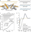
Figure 1
Figure 1
(a) Model of two identical TAV2b fragments (aa 21–53, sTAV2b, orange) bound to duplex RNA (grey). Model was derived from crystal structure PDB ID: 2ZI0. (b) Oligonucleotide sequences used to test duplex binding (RNA: gray, DNA: blue). (c) Representative ITC plots of complex formation between sTAV2b (c=108 μM) and RNAα/DNAβ (c(duplex)=6 μM). Measurements were performed in triplicate, errors=1σ; for full data see Supporting Information Table S4 and Figure S4. Buffer: Phosphate‐buffered saline (pH 7.4) (d) CD spectra of sTAV2b (c=4 μM; orange line), RNAα/DNAβ (c(duplex)=2  μM; blue line), spectra of RNAα/DNAβ (c(duplex)=2 μM) with sTAV2b (c=4 μM) (black line) and the sum of the two individual spectra (dotted black line). Buffer: 10 mM sodium phosphate (pH 7.4), 100 mM NaCl.
Figure 2
Figure 2
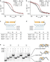
(a) Schematic overview of the design of conjugate type A and B: sTAV2b (orange) bound to duplex RNA/DNA (grey/blue) serves as starting point for the design of peptide‐DNA conjugates (1‐A12 and 2‐B12) targeting single stranded RNAα (grey). Chemical structure of linkages between peptide and DNA are shown. (b) EMSA of RNAα in the presence and absence of DNA A12, a mixture of DNA and peptide (A12+1) and conjugate 1‐A12. Experiments employed 15 % native polyacrylamide gel electrophoresis (PAGE) (c(RNA)=3 μM, c(ligand)=4.5 μM. Running buffer: 1x TAE, stain: SYBR gold (full gel in Supporting Information Figure S8). (c) K d‐values determined by ITC for RNAα‐ligand interactions (triplicate measurements, errors=1σ; for full data see Supporting Information Table S4 and Figures S9‐11). (d) Representative ITC of 1‐A12 with the RNAα (for full data see Supporting Information Figure S11). (e) EMSA of RNAα in the presence and absence of DNA B12, a mixture of DNA and peptide (B12 + 2) and conjugate 2‐B12 (for details see caption Figure 2b, full gel in Supporting Information Figure S12). (f) K d‐values determined by ITC for RNAα/ligand interactions (triplicate measurements, errors=1σ; for full data see Supporting Information Table S4 and Figures S13–15).(g) Representative ITC of 2‐B12 with the RNAα (for all data see Supporting Information Figure S15).
Figure 3
Figure 3
(a) Melting temperature profiles of RNAα in the presence of A12, an equimolar mixture of A12+1, and 1‐A12 respectively. (b) Melting temperature profiles of RNAα in the presence of B12, an equimolar mixture of B12+2, and 2‐B12 respectively (λ=267 nm, c(RNAα)=2 μM, c(binding partners)=2 μM. Buffer: 10 mM sodium phosphate, 100 mM NaCl (pH 7.4). (c) Sequences of DNA‐truncated A‐family conjugates. Table of T m‐values of conjugates with RNAα (for melting curves see Supporting Information Figure S18). (d) Sequences of DNA‐truncated B‐ family conjugates. Table of T m‐values of conjugates with RNAα (for melting curves see Supporting Information Figure S18). (e) EMSA of RNAα in the presence of different conjugates (Supporting Information Figure S19) including co‐incubation of A‐ and B‐series members with DNA sequences of equal lengths. Experiments employed 15 % native PAGE (c(RNA)=3 μM, c(conjugate)=4.5 μM. Running buffer: TAE, stain: SYBR gold. Cartoon representations of the proposed RNA/conjugate complexes corresponding to band species are presented on the right hand side.
Figure 4
Figure 4
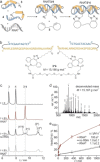
(a) Schematic overview of RNAα‐templated reaction. Azide‐modified conjugates 3 and 4 bind RNAα to form a ternary complex which reacts with a bis‐alkyne crosslinker to yield a complex between ligated conjugate 3*4 and RNAα. (b) Structure of heterodimeric product 3*4 including the chemical structure of the reacted crosslink. (c) HPLC traces of unreacted 3 and 4 (t=0) and of reactions between 3, 4 and 5 after 6 h in the presence of RNAα (red line), in the absence of RNA (no RNA), and in the presence of RNAβ (gray). (d) Electrospray ionization (ESI) mass spectrum of heterodimeric product 3*4 (full characterization see Supporting Information Figure S23). (e) Time‐dependent yields of 3*4 as determined by HPLC including initial rates (v r). Conjugates (c=5 μM) were incubated for 18 h at room temperature in the presence or absence of an RNA template (c=5 μM) and bis‐alkyne crosslinker 5 (c=5.75 μM). Reaction buffer: 100 mM NH4HCO3, 20 % acetonitrile, 1 % DMSO (pH=8.0). Templated reactions were performed in triplicate, errors=1σ; for full data see Supporting Information Table S6 and Figure S27.

Similar articles

References

    1. J. Wilce, J. Vivian, M. Wilce, in Protein Dimerization and Oligomerization in Biology (Ed. J. M. Matthews), Springer, New York, 2012, pp. 91–104.
    1. Cassandri M., Smirnov A., Novelli F., Pitolli C., Agostini M., Malewicz M., Melino G., Raschellà G., Cell Death Dis. 2017, 3, 17071. - PMC - PubMed
    1. Jones S., Genome Biol. 2004, 5, 226. - PMC - PubMed
    1. Zhao D., Kong Y., Zhao S., Xing H., Top. Curr. Chem. 2020, 378, 41. - PubMed
    1. Dovgan I., Koniev O., Kolodych S., Wagner A., Bioconjugate Chem. 2019, 30, 2483–2501. - PubMed