. 2022 Feb 21;118(3):772-784.
doi: 10.1093/cvr/cvab156.

Drebrin attenuates atherosclerosis by limiting smooth muscle cell transdifferentiation

Affiliations

Drebrin attenuates atherosclerosis by limiting smooth muscle cell transdifferentiation

Jiao-Hui Wu et al. Cardiovasc Res. .

Abstract

Aims: The F-actin-binding protein Drebrin inhibits smooth muscle cell (SMC) migration, proliferation, and pro-inflammatory signalling. Therefore, we tested the hypothesis that Drebrin constrains atherosclerosis.

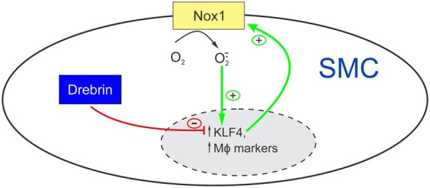
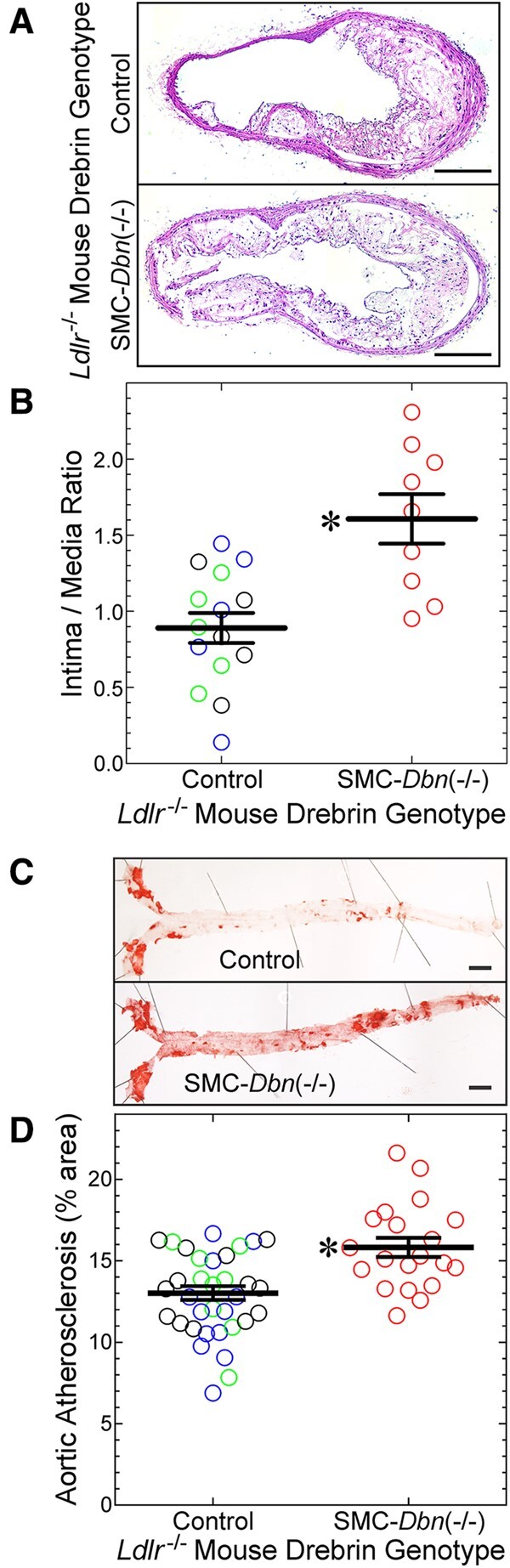
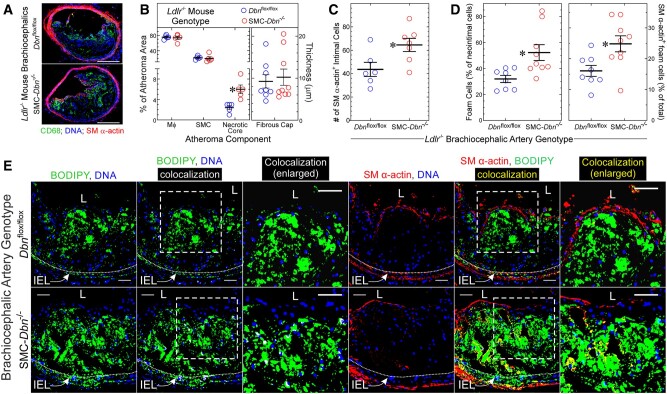
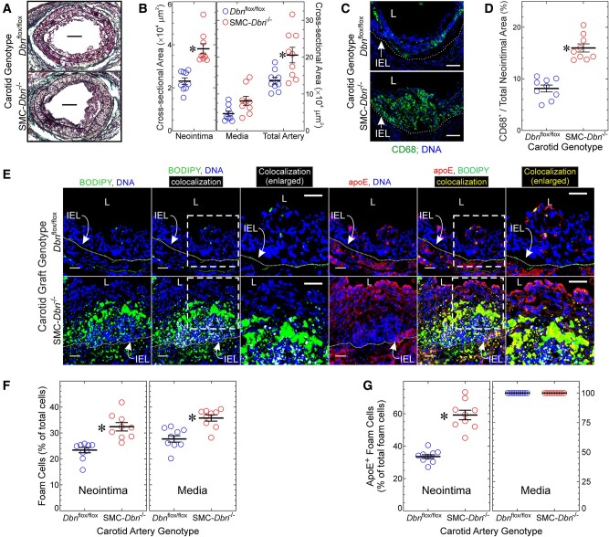
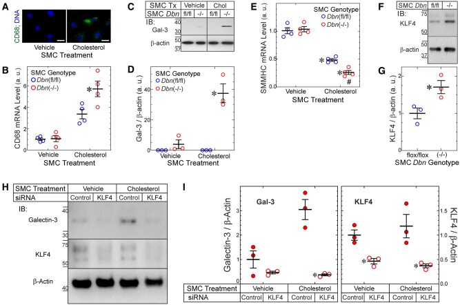
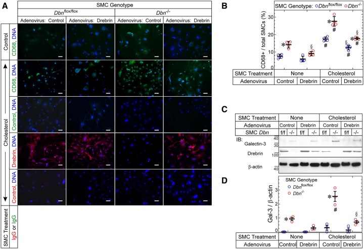
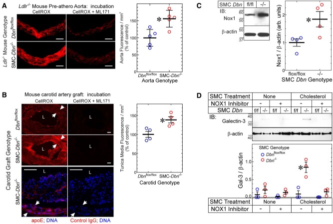
Methods and results: SM22-Cre+/Dbnflox/flox/Ldlr-/- (SMC-Dbn-/-/Ldlr-/-) and control mice (SM22-Cre+/Ldlr-/-, Dbnflox/flox/Ldlr-/-, and Ldlr-/-) were fed a western diet for 14-20 weeks. Brachiocephalic arteries of SMC-Dbn -/-/Ldlr-/- mice exhibited 1.5- or 1.8-fold greater cross-sectional lesion area than control mice at 14 or 20 weeks, respectively. Aortic atherosclerotic lesion surface area was 1.2-fold greater in SMC-Dbn-/-/Ldlr-/- mice. SMC-Dbn-/-/Ldlr-/- lesions comprised necrotic cores that were two-fold greater in size than those of control mice. Consistent with their bigger necrotic core size, lesions in SMC-Dbn-/- arteries also showed more transdifferentiation of SMCs to macrophage-like cells: 1.5- to 2.5-fold greater, assessed with BODIPY or with CD68, respectively. In vitro data were concordant: Dbn-/- SMCs had 1.7-fold higher levels of KLF4 and transdifferentiated to macrophage-like cells more readily than Dbnflox/flox SMCs upon cholesterol loading, as evidenced by greater up-regulation of CD68 and galectin-3. Adenovirally mediated Drebrin rescue produced equivalent levels of macrophage-like transdifferentiation in Dbn-/- and Dbnflox/flox SMCs. During early atherogenesis, SMC-Dbn-/-/Ldlr-/- aortas demonstrated 1.6-fold higher levels of reactive oxygen species than control mouse aortas. The 1.8-fold higher levels of Nox1 in Dbn-/- SMCs were reduced to WT levels with KLF4 silencing. Inhibition of Nox1 chemically or with siRNA produced equivalent levels of macrophage-like transdifferentiation in Dbn-/- and Dbnflox/flox SMCs.

Conclusion: We conclude that SMC Drebrin limits atherosclerosis by constraining SMC Nox1 activity and SMC transdifferentiation to macrophage-like cells.

Keywords: Atherosclerosis; Drebrin; Foam cell; NADPH oxidase; Nox1; Reactive oxygen species; VSMC; Vascular smooth muscle cells.


Figures

None
Graphical abstract
Figure 1
Figure 1
SMC Drebrin attenuates atherosclerosis. The indicated male mice were fed a western diet for 20 weeks, from age 8 weeks: SMC-Dbn//Ldlr/ mice, and ‘control’ mice [SM22-Cre+/Ldlr/ (green), Dbnflox/flox/Ldlr/ (blue), and Dbn+/+/Ldlr/ (black)]. All control groups yielded congruent data; therefore, data from these groups were pooled. (A) Brachiocephalic artery sections from the indicated mice were stained with H&E; one specimen of each group is shown, representing 9–15 examined per group. Scale bars = 200 μm. (B) Brachiocephalic cross-sectional neointimal and medial areas were measured by planimetry for control (n = 15) and SMC-Dbn//Ldlr/ mice (n = 9). For each artery, neointimal area was divided by medial area; these ratios for each mouse were plotted along with group means ± S.E. Compared with control mice: *P <0.001 by t test. (C) Aortas from these mice were excised from the root to the iliac bifurcation, stained with Sudan IV, pinned and photographed en face. Scale bars = 2 mm. (D) The percentage of aortic surface area comprising Sudanophilic lesions was quantitated and plotted for each mouse [control (n = 33) and SMC-Dbn//Ldlr/ (n = 20)], along with group means ± SE. Compared with control: *P <0.001 (t test).
Figure 2
Figure 2
SMC Drebrin deficiency augments atherosclerotic lesion necrotic core size and the prevalence of SMC-derived foam cells. (A) Brachiocephalic arteries from Dbnflox/flox/Ldlr/ (control) and SMC-Dbn//Ldlr/ mice used in Figure 1 were immunostained with IgG specific for macrophages (CD68, green) and smooth muscle (SM) α-actin (red); all specimens were counterstained for DNA (blue). Serial sections stained with isotype control IgG yielded no green or red colour (not shown). Scale bars = 200 µm. (B) The percentages of atheroma area comprising macrophages, SMCs, and necrotic core were measured with ImageJ for five discrete mice of each group. The thickness of the atheroma SM α-actin+ fibrous cap was measured at six locations, which were averaged and plotted for each artery (n = 8–9), along with means ± SE for each genotype. Compared with control: *P <0.01 (multiple t tests, Holm–Sidak correction for multiple comparisons). (C) SM α-actin-positive cells in each brachiocephalic atheroma were counted manually, and plotted with mean ± SE of 6–7 mice/genotype. Compared with control: *P <0.03 (t test). (D, E) Serial sections of brachiocephalic arteries from A and B were incubated simultaneously with BODIPY® 493/503 (for cholesteryl ester), Hoechst 33342 (DNA), and either Cy3-conjugated IgG specific for SM α-actin or for no known protein. Confocal microscopy used an optical slice thickness of 1 µm. Serial sections stained with Cy3-control IgG yielded no red colour (not shown). The dotted white lines indicate the internal elastic lamina (IEL). The dashed boxes indicate areas enlarged further in the adjacent panels. L, lumen. Scale bars = 50 µm. Co-localization of green (BODIPY) with either blue (Hoechst) or red (SM α-actin) was performed using Imaris 9.2 software, to yield white or yellow, respectively. BODIPY-stained material in the neointima or media was judged to be cellular (as opposed to extracellular), and therefore foam cells, if there was co-localization of green BODIPY with blue DNA fluorescence (designated white). (D) Within several neointimal microscopic fields, the number of BODIPY+ (foam) cells (≥100 per artery) was divided by the total number of cells to obtain neointimal foam cell prevalence; the number of foam cells showing yellow SM α-actin co-localization was divided by the total number of neointimal foam cells to obtain ‘% of total foam cells’. Data were plotted for distinct brachiocephalic arteries from control (n = 8) and SMC-Dbn//Ldlr/ mice (n = 9), along with means ± SE. Compared with control: *P <0.02 (t test).
Figure 3
Figure 3
SMC Drebrin inhibits atherosclerosis and SMC-to-foam cell transdifferentiation. Common carotid arteries from Dbnflox/flox and SMC-Dbn/ mice were transplanted into the right common carotid of congenic Apoe/ mice as interposition grafts, and harvested 4 weeks later. (A) Sections were stained with a modified connective tissue stain to facilitate planimetry of neointima and media. Scale bars = 100 μm. (B) Neointimal, medial, and total arterial cross-sectional areas were measured by planimetry (ImageJ), and plotted for nine distinct carotid grafts/genotype along with means ± SE. Compared with Dbnflox/flox: *P <0.01 (Mann–Whitney). (C) Serial sections of carotid grafts from A were incubated with anti-CD68 or isotype control IgG, along with Hoechst 33342 (DNA). Negative control IgG yielded no colour (not shown). Scale bars = 50 μm. L, lumen; IEL, internal elastic lamina. (D) In cross sections from C, the CD68-positive neointimal area was divided by the cognate total neointimal area, and plotted for nine distinct carotid grafts/genotype, along with means ± SE. Compared with Dbnflox/flox: *P <10−3 (t test). (E) Serial sections of carotid grafts were incubated simultaneously with BODIPY® 493/503 and goat anti-apoE, followed by Hoechst 33342 (DNA) and anti-goat/Alexa 546 IgG. Confocal microscopy used an optical slice thickness of 1 μm. Serial sections stained with non-immune primary IgG yielded no colour (not shown). The dotted white lines indicate the IEL. The dashed boxes indicate areas enlarged further in the adjacent panels. L, lumen. Scale bars = 20 μm. Co-localization of red (apoE) with either blue (Hoechst) or green (BODIPY) was performed as in Figure 2. (F) BODIPY-stained material was judged to be cellular as in Figure 2. Foam cell prevalence was determined as in Figure 2 and plotted for 9 distinct carotids/genotype, along with means ± SE. Compared with Dbnflox/flox: *P <10−3 (Mann–Whitney). (G) BODIPY+ neointimal or medial cells (≥100 per layer per carotid graft) were scored as containing yellow (apoE+/BODIPY+) or not; the percentage of apoE+/BODIPY+ in each layer was plotted for nine distinct carotids per genotype, along with means ± SE. Compared with Dbnflox/flox: *P <10−3 (t test).
Figure 4
Figure 4
Drebrin inhibits SMC-to-foam cell transdifferentiation in a KLF4-dependent manner. (A) WT SMCs were treated with vehicle (control) or cholesterol-methyl-β-cyclodextrin (10 µg/mL) for 72 h and stained with anti-CD68 IgG. (Isotype control IgG yielded no colour.) Scale bar = 10 µm. Results represent three independent experiments with >100 SMCs evaluated per experiment. (B) Dbnflox/flox and SMC-Dbn/ SMCs were treated ± cholesterol (as in A) for 24 h before mRNA isolation and qRT-PCR for CD68 and GAPDH. Threshold CD68 counts were normalized to cognate GAPDH values and plotted from four experiments (means ± SE) using three sets of independently isolated Dbnflox/flox and SMC-Dbn/ SMC lines. Compared with cognate Dbnflox/flox: *P <0.05 (two-way ANOVA with Sidak post hoc test). (C) SMCs from B were treated (Tx) as above with vehicle or cholesterol (‘Chol’) for 48 h; soluble SMC extracts were immunoblotted sequentially for Galectin-3 (Gal-3) and β-actin. (D) Band densities for Gal-3 were normalized to cognate β-actin band densities; ratios were plotted from three independent experiments (means ± SE) with distinct pairs of Dbnflox/flox and SMC-Dbn/ SMC lines. Compared with vehicle control: *P <0.01 (two-way ANOVA with Sidak post hoc test). (E) The mRNA of SMCs from B was subjected to qRT-PCR for SMMHC and GAPDH; data were obtained and processed as in B. Compared with vehicle-treated SMCs: *P <0.001; compared with Dbnflox/flox: #, P <0.05 (two-way ANOVA with Sidak post hoc test, n = 4/group). (F) Dbnflox/flox and SMC-Dbn/ SMCs were grown to confluence, serum starved for 24 h, and then solubilized. SMC lysates were immunoblotted sequentially for KLF4 and β-actin. KLF migrates as a doublet due to its known post-translational modifications. (G) Band densities for KLF4 were normalized to cognate β-actin band densities; the ratios were plotted as arbitrary units (a. u.) from 3 independent experiments with three sets of independently isolated Dbnflox/flox and SMC-Dbn/ SMC lines. Compared with Dbnflox/flox: *P <0.05 (t test). (H) SMC-Dbn/ SMCs were transfected with siRNAs targeting KLF4 or no known mRNA (‘control’). Forty-eight hours later, SMCs were treated for a further 48 h with cholesterol-methyl-β-cyclodextrin (10 µg/mL, ‘cholesterol’) or vehicle, and then solubilized. SMC lysates were immunoblotted sequentially for Galectin-3, KLF4 and β-actin. (I) Band densities for Gal-3 or KLF4 were normalized to cognate β-actin band densities; the ratios were plotted as means ± SE from three independent experiments with two independently isolated primary SMC-Dbn/ SMC lines. Compared with cognate control siRNA-transfected SMCs: *P <0.01 (Gal-3) or P <0.05 (KLF4), two-way ANOVA with Sidak post hoc test.
Figure 5
Figure 5
Drebrin inhibits SMC up-regulation of CD68 and Galectin-3. (A) Aortic SMCs from Dbnflox/flox and SMC-Dbn/ mice were transduced with empty vector or Drebrin-encoding adenoviruses and then exposed to medium containing vehicle or solubilized cholesterol for 48 h (37°C). SMCs were then immunostained with IgG specific for CD68 or Drebrin (or isotype control IgG), as indicated, counter-stained for DNA, and imaged at 200× (original magnification). Scale bars = 50 μm. (B) For each SMC group, the number of CD68+ SMCs was divided by the total number of SMCs and multiplied by 100; the resulting percentages are plotted (along with means ± SE) for four experiments with four distinct pairs of Dbnflox/flox and Dbn/ SMCs. P <0.05 for comparisons of (i) Dbn/ vs. cognate Dbnflox/flox SMCs (*); (ii) Drebrin adenovirus-transduced vs. cognate control adenovirus-transduced SMCs (§); (iii) cholesterol-treated vs. vehicle-treated SMCs (#) (two-way ANOVA with Sidak post hoc test). (C) Dbnflox/flox (f/f) and Dbn/ SMCs were transduced and treated ± cholesterol just as in A. After the 48-h incubation ± cholesterol; however, SMCs were solubilized; protein extracts were immunoblotted serially for the indicated proteins. (D) The band intensities for Galectin-3 were normalized to cognate β-actin band densities, and these ratios were plotted (along with means ± SE) for three experiments with three distinct pairs of Dbnflox/flox and Dbn/ SMCs. P <0.05 for comparisons of (i) Dbn/ vs. cognate Dbnflox/flox SMCs (*); (ii) cholesterol-treated vs. vehicle-treated SMCs (#); (iii) Drebrin adenovirus-transduced vs. cognate control adenovirus-transduced SMCs (§) (two-way ANOVA with Sidak post hoc test).
Figure 6
Figure 6
Drebrin regulates SMC ROS levels and foam cell transdifferentiation via Nox1. (A) Serial frozen sections of Dbnflox/flox/Ldlr/ and SMC-Dbn//Ldlr/ pre-atherosclerotic aortas used in Supplementary material online, Figure S2 were incubated without (negative control) or with CellROX® Orange in the absence (total signal) or presence (nonspecific, Nox1-independent signal) of the Nox1 inhibitor ML171 (see Section 2). Fluorescence photomicrographs from single aortas of each genotype are shown; negative control samples yielded no red fluorescence (not shown). Scale bars = 50 μm. (Right), Specific CellROX® fluorescence was calculated as the fluorescence (red pixels/mm2) in aortic slices incubated with CellROX® minus that obtained from slices incubated with CellROX® plus ML171; values from five distinct aortas of each genotype were plotted, along with means ± SE. Compared with Dbnflox/flox/Ldlr/: *P <0.01 (t test). (B) Frozen sections from atherosclerotic carotid interposition grafts used in Figure 3 were processed like the aortas of A; serial sections were immunostained with goat IgG specific for apoE or for no known protein (control). ‘L’, lumen. Arrows indicate the internal and external elastic laminae (and thus the tunica media). Scale bars = 50 μm. (Right) Specific CellROX® fluorescence in the tunica media was quantitated as in B. Values for four discrete carotid grafts per genotype are plotted, with means ± SE. Compared with Dbnflox/flox carotid grafts: *P <0.03 (Mann–Whitney test). (C) SMCs from age- and sex-matched Dbn/ and Dbnflox/flox (fl/fl) mice were solubilized and immunoblotted serially for Nox1 and β-actin. Nox1 band densities were normalized to cognate β-actin band densities; ratios were plotted as arbitrary units for four independent pairs of Dbnflox/flox and Dbn/ SMC lines, with means ± SE. Compared with Dbnflox/flox: *P <0.03 (Mann–Whitney test). (D) Dbn/ and Dbnflox/flox (‘f/f’) SMCs were treated without or with the Nox1-selective inhibitor ML171 (1 μmol/L) in the presence or absence of cholesterol-methyl-β-cyclodextrin (10 µg/mL) or vehicle for 24 h and then solubilized. SMC lysates were immunoblotted sequentially for Galectin-3 and β-actin. Band densities for galectin-3 were normalized to cognate β-actin band densities; the ratios were plotted as means ± SE from three independent experiments with two independently isolated SMC lines of each genotype. Compared with cognate Dbnflox/flox SMCs: *P <0.05 (two-way ANOVA with Sidak post hoc test).

References

    1. Wang N, Tabas I, Winchester R, Ravalli S, Rabbani LE, Tall A.. Interleukin 8 is induced by cholesterol loading of macrophages and expressed by macrophage foam cells in human atheroma. J Biol Chem 1996;271:8837–8842. - PubMed
    1. Galis ZS, Sukhova GK, Kranzhofer R, Clark S, Libby P.. Macrophage foam cells from experimental atheroma constitutively produce matrix-degrading proteinases. Proc Natl Acad Sci U S A 1995;92:402–406. - PMC - PubMed
    1. Childs BG, Baker DJ, Wijshake T, Conover CA, Campisi J, van Deursen JM.. Senescent intimal foam cells are deleterious at all stages of atherosclerosis. Science 2016;354:472–477. - PMC - PubMed
    1. Rong JX, Shapiro M, Trogan E, Fisher EA.. Transdifferentiation of mouse aortic smooth muscle cells to a macrophage-like state after cholesterol loading. Proc Natl Acad Sci U S A 2003;100:13531–13536. - PMC - PubMed
    1. Vengrenyuk Y, Nishi H, Long X, Ouimet M, Savji N, Martinez FO, Cassella CP, Moore KJ, Ramsey SA, Miano JM, Fisher EA.. Cholesterol loading reprograms the microRNA-143/145-myocardin axis to convert aortic smooth muscle cells to a dysfunctional macrophage-like phenotype. Arterioscler Thromb Vasc Biol 2015;35:535–546. - PMC - PubMed

Publication types